1IDO

I-DOMAIN FROM INTEGRIN CR3, MG2+ BOUND


Experimental Data Snapshot

  • Method: X-RAY DIFFRACTION
  • Resolution: 1.70 Å
  • R-Value Work: 0.202 
  • R-Value Observed: 0.202 

wwPDB Validation   3D Report Full Report


This is version 1.4 of the entry. See complete history


Literature

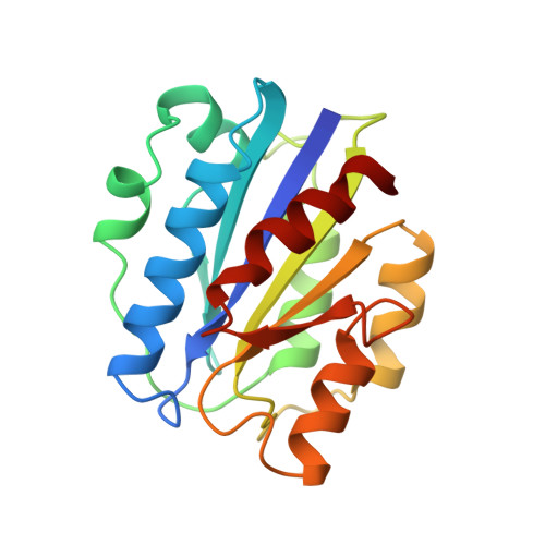
Crystal structure of the A domain from the alpha subunit of integrin CR3 (CD11b/CD18).

Lee, J.O.Rieu, P.Arnaout, M.A.Liddington, R.

(1995) Cell 80: 631-638

  • DOI: https://doi.org/10.1016/0092-8674(95)90517-0
  • Primary Citation of Related Structures:  
    1IDO

  • PubMed Abstract: 

    We have determined the high resolution crystal structure of the A domain from the alpha chain of integrin CR3. The domain adopts a classic alpha/beta "Rossmann" fold and contains an unusual Mg2+ coordination site at its surface. One of the coordinating ligands is the glutamate side chain from another A domain molecule. We suggest that this site represents a general metal ion-dependent adhesion site (MIDAS) for binding protein ligands. We further propose that the beta subunits of integrins contain a MIDAS motif within a modified A domain. Our crystal structure will allow reliable models to be built for other members of the A domain superfamily and should facilitate development of novel adhesion modulatory drugs.


  • Organizational Affiliation

    Laboratory of X-Ray Crystallography, Dana Farber Cancer Institute, Harvard Medical School Boston, Massachusetts 02115.


Macromolecules
Find similar proteins by:  (by identity cutoff)  |  3D Structure
Entity ID: 1
MoleculeChains Sequence LengthOrganismDetailsImage
INTEGRIN189Homo sapiensMutation(s): 0 
UniProt & NIH Common Fund Data Resources
Find proteins for P11215 (Homo sapiens)
Explore P11215 
Go to UniProtKB:  P11215
PHAROS:  P11215
GTEx:  ENSG00000169896 
Entity Groups  
Sequence Clusters30% Identity50% Identity70% Identity90% Identity95% Identity100% Identity
UniProt GroupP11215
Sequence Annotations
Expand
  • Reference Sequence
Small Molecules
Ligands 1 Unique
IDChains Name / Formula / InChI Key2D Diagram3D Interactions
MG
Query on MG

Download Ideal Coordinates CCD File 
B [auth A]MAGNESIUM ION
Mg
JLVVSXFLKOJNIY-UHFFFAOYSA-N
Experimental Data & Validation

Experimental Data

  • Method: X-RAY DIFFRACTION
  • Resolution: 1.70 Å
  • R-Value Work: 0.202 
  • R-Value Observed: 0.202 
  • Space Group: P 43
Unit Cell:
Length ( Å )Angle ( ˚ )
a = 45.7α = 90
b = 45.7β = 90
c = 94.3γ = 90
Software Package:
Software NamePurpose
X-PLORmodel building
X-PLORrefinement
DENZOdata reduction
SCALEPACKdata scaling
X-PLORphasing

Structure Validation

View Full Validation Report


Entry History 

Deposition Data

Revision History  (Full details and data files)

  • Version 1.0: 1996-08-01
    Type: Initial release
  • Version 1.1: 2008-03-24
    Changes: Version format compliance
  • Version 1.2: 2011-07-13
    Changes: Version format compliance
  • Version 1.3: 2018-03-21
    Changes: Data collection, Other
  • Version 1.4: 2024-02-07
    Changes: Data collection, Database references, Derived calculations