1IC2

DECIPHERING THE DESIGN OF THE TROPOMYOSIN MOLECULE


Experimental Data Snapshot

  • Method: X-RAY DIFFRACTION
  • Resolution: 2.00 Å
  • R-Value Free: 0.283 
  • R-Value Work: 0.249 

wwPDB Validation   3D Report Full Report


This is version 1.4 of the entry. See complete history


Literature

Deciphering the design of the tropomyosin molecule

Brown, J.H.Kim, K.-H.Jun, G.Greenfield, N.J.Dominguez, R.Volkmann, N.Hitchcock-DeGregori, S.E.Cohen, C.

(2001) Proc Natl Acad Sci U S A 98: 8496-8501

  • DOI: https://doi.org/10.1073/pnas.131219198
  • Primary Citation of Related Structures:  
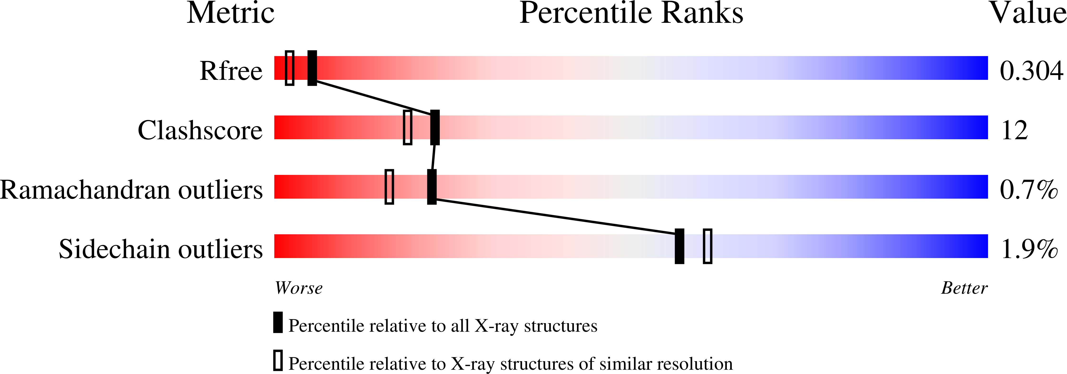
    1IC2

  • PubMed Abstract: 

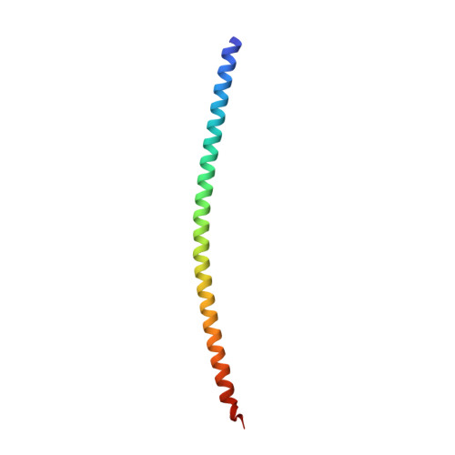
    The crystal structure at 2.0-A resolution of an 81-residue N-terminal fragment of muscle alpha-tropomyosin reveals a parallel two-stranded alpha-helical coiled-coil structure with a remarkable core. The high alanine content of the molecule is clustered into short regions where the local 2-fold symmetry is broken by a small (approximately 1.2-A) axial staggering of the helices. The joining of these regions with neighboring segments, where the helices are in axial register, gives rise to specific bends in the molecular axis. We observe such bends to be widely distributed in two-stranded alpha-helical coiled-coil proteins. This asymmetric design in a dimer of identical (or highly similar) sequences allows the tropomyosin molecule to adopt multiple bent conformations. The seven alanine clusters in the core of the complete molecule (which spans seven monomers of the actin helix) promote the semiflexible winding of the tropomyosin filament necessary for its regulatory role in muscle contraction.


  • Organizational Affiliation

    Rosenstiel Basic Medical Sciences Research Center, Brandeis University, Waltham, MA 02454-9110, USA.


Macromolecules
Find similar proteins by:  (by identity cutoff)  |  3D Structure
Entity ID: 1
MoleculeChains Sequence LengthOrganismDetailsImage
TROPOMYOSIN ALPHA CHAIN, SKELETAL MUSCLE
A, B, C, D
81Gallus gallusMutation(s): 1 
UniProt
Find proteins for P04268 (Gallus gallus)
Explore P04268 
Go to UniProtKB:  P04268
Entity Groups  
Sequence Clusters30% Identity50% Identity70% Identity90% Identity95% Identity100% Identity
UniProt GroupP04268
Sequence Annotations
Expand
  • Reference Sequence
Experimental Data & Validation

Experimental Data

  • Method: X-RAY DIFFRACTION
  • Resolution: 2.00 Å
  • R-Value Free: 0.283 
  • R-Value Work: 0.249 
  • Space Group: P 1
Unit Cell:
Length ( Å )Angle ( ˚ )
a = 41.4α = 93.7
b = 45.5β = 98.1
c = 56.3γ = 104.4
Software Package:
Software NamePurpose
MLPHAREphasing
CNSrefinement
DENZOdata reduction
CCP4data scaling

Structure Validation

View Full Validation Report


Entry History 

Deposition Data

Revision History  (Full details and data files)

  • Version 1.0: 2001-07-25
    Type: Initial release
  • Version 1.1: 2008-04-27
    Changes: Version format compliance
  • Version 1.2: 2011-07-13
    Changes: Version format compliance
  • Version 1.3: 2021-10-27
    Changes: Database references
  • Version 1.4: 2024-02-07
    Changes: Data collection