1I9V

CRYSTAL STRUCTURE ANALYSIS OF A TRNA-NEOMYCIN COMPLEX


Experimental Data Snapshot

  • Method: X-RAY DIFFRACTION
  • Resolution: 2.60 Å
  • R-Value Free: 0.275 
  • R-Value Work: 0.228 
  • R-Value Observed: 0.230 

wwPDB Validation   3D Report Full Report


This is version 1.5 of the entry. See complete history


Literature

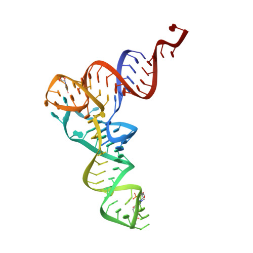
Aminoglycoside binding displaces a divalent metal ion in a tRNA-neomycin B complex.

Mikkelsen, N.E.Johansson, K.Virtanen, A.Kirsebom, L.A.

(2001) Nat Struct Biol 8: 510-514

  • DOI: https://doi.org/10.1038/88569
  • Primary Citation of Related Structures:  
    1I9V

  • PubMed Abstract: 

    Aminoglycosides bind to RNA and interfere with its function, and it has been suggested that aminoglycoside binding to RNA displaces essential divalent metal ions. Here we demonstrate that addition of various aminoglycosides inhibited Pb2+-induced cleavage of yeast tRNA(Phe). Cocrystallization of yeast tRNA(Phe) and an aminoglycoside, neomycin B, resulted in crystals that diffracted to 2.6 A and the structure of the complex was solved by molecular replacement. The structure shows that the neomycin B binding site overlaps with known divalent metal ion binding sites in yeast tRNA(Phe), providing direct evidence for the hypothesis that aminoglycosides displace metal ions. Additionally, the neomycin B binding site overlaps with major determinants for Escherichia coli phenylalanyl-tRNA-synthetase. Here we present data demonstrating that addition of neomycin B inhibited aminoacylation of E. coli tRNA(Phe) in the mid microM range. Given that aminoglycoside and metal ion binding sites overlap, we discuss that aminoglycosides can be considered as 'metal mimics'.


  • Organizational Affiliation

    Department of Cell and Molecular Biology, Uppsala University, Box 596, Biomedical Center, SE-751 24, Uppsala, Sweden.


Macromolecules
Find similar nucleic acids by:  (by identity cutoff)  |  3D Structure
Entity ID: 1
MoleculeChains LengthOrganismImage
PHENYLALANINE TRANSFER RNA76Saccharomyces cerevisiae
Sequence Annotations
Expand
  • Reference Sequence
Small Molecules
Binding Affinity Annotations 
IDSourceBinding Affinity
NMY PDBBind:  1I9V Ki: 3.42e+5 (nM) from 1 assay(s)
Experimental Data & Validation

Experimental Data

  • Method: X-RAY DIFFRACTION
  • Resolution: 2.60 Å
  • R-Value Free: 0.275 
  • R-Value Work: 0.228 
  • R-Value Observed: 0.230 
  • Space Group: P 1 21 1
Unit Cell:
Length ( Å )Angle ( ˚ )
a = 54.9α = 90
b = 32.9β = 90.5
c = 63.1γ = 90
Software Package:
Software NamePurpose
AMoREphasing
CNSrefinement
DENZOdata reduction
SCALEPACKdata scaling

Structure Validation

View Full Validation Report


Entry History 

Deposition Data

Revision History  (Full details and data files)

  • Version 1.0: 2001-06-04
    Type: Initial release
  • Version 1.1: 2008-04-27
    Changes: Version format compliance
  • Version 1.2: 2011-07-13
    Changes: Version format compliance
  • Version 1.3: 2018-03-07
    Changes: Data collection
  • Version 1.4: 2022-12-21
    Changes: Database references, Derived calculations
  • Version 1.5: 2023-09-20
    Changes: Data collection, Refinement description