1I9B

X-RAY STRUCTURE OF ACETYLCHOLINE BINDING PROTEIN (ACHBP)


Experimental Data Snapshot

  • Method: X-RAY DIFFRACTION
  • Resolution: 2.70 Å
  • R-Value Free: 0.297 
  • R-Value Work: 0.260 
  • R-Value Observed: 0.260 

wwPDB Validation   3D Report Full Report


This is version 1.3 of the entry. See complete history


Literature

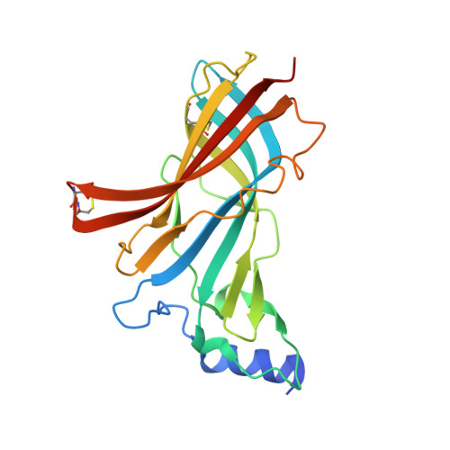
Crystal structure of an ACh-binding protein reveals the ligand-binding domain of nicotinic receptors.

Brejc, K.van Dijk, W.J.Klaassen, R.V.Schuurmans, M.van Der Oost, J.Smit, A.B.Sixma, T.K.

(2001) Nature 411: 269-276

  • DOI: https://doi.org/10.1038/35077011
  • Primary Citation of Related Structures:  
    1I9B

  • PubMed Abstract: 

    Pentameric ligand gated ion-channels, or Cys-loop receptors, mediate rapid chemical transmission of signals. This superfamily of allosteric transmembrane proteins includes the nicotinic acetylcholine (nAChR), serotonin 5-HT3, gamma-aminobutyric-acid (GABAA and GABAC) and glycine receptors. Biochemical and electrophysiological information on the prototypic nAChRs is abundant but structural data at atomic resolution have been missing. Here we present the crystal structure of molluscan acetylcholine-binding protein (AChBP), a structural and functional homologue of the amino-terminal ligand-binding domain of an nAChR alpha-subunit. In the AChBP homopentamer, the protomers have an immunoglobulin-like topology. Ligand-binding sites are located at each of five subunit interfaces and contain residues contributed by biochemically determined 'loops' A to F. The subunit interfaces are highly variable within the ion-channel family, whereas the conserved residues stabilize the protomer fold. This AChBP structure is relevant for the development of drugs against, for example, Alzheimer's disease and nicotine addiction.


  • Organizational Affiliation

    Division of Molecular Carcinogenesis, Netherlands Cancer Institute, Plesmanlaan 121, 1066 CX Amsterdam, The Netherlands.


Macromolecules
Find similar proteins by:  (by identity cutoff)  |  3D Structure
Entity ID: 1
MoleculeChains Sequence LengthOrganismDetailsImage
ACETYLCHOLINE BINDING PROTEIN
A, B, C, D, E
217Lymnaea stagnalisMutation(s): 0 
UniProt
Find proteins for P58154 (Lymnaea stagnalis)
Explore P58154 
Go to UniProtKB:  P58154
Entity Groups  
Sequence Clusters30% Identity50% Identity70% Identity90% Identity95% Identity100% Identity
UniProt GroupP58154
Sequence Annotations
Expand
  • Reference Sequence
Small Molecules
Ligands 2 Unique
IDChains Name / Formula / InChI Key2D Diagram3D Interactions
EPE
Query on EPE

Download Ideal Coordinates CCD File 
H [auth A],
J [auth B],
L [auth C],
N [auth D],
R [auth E]
4-(2-HYDROXYETHYL)-1-PIPERAZINE ETHANESULFONIC ACID
C8 H18 N2 O4 S
JKMHFZQWWAIEOD-UHFFFAOYSA-N
CA
Query on CA

Download Ideal Coordinates CCD File 
F [auth A]
G [auth A]
I [auth B]
K [auth C]
M [auth D]
F [auth A],
G [auth A],
I [auth B],
K [auth C],
M [auth D],
O [auth E],
P [auth E],
Q [auth E]
CALCIUM ION
Ca
BHPQYMZQTOCNFJ-UHFFFAOYSA-N
Experimental Data & Validation

Experimental Data

  • Method: X-RAY DIFFRACTION
  • Resolution: 2.70 Å
  • R-Value Free: 0.297 
  • R-Value Work: 0.260 
  • R-Value Observed: 0.260 
  • Space Group: P 42 21 2
Unit Cell:
Length ( Å )Angle ( ˚ )
a = 141.66α = 90
b = 141.66β = 90
c = 120.87γ = 90
Software Package:
Software NamePurpose
DENZOdata reduction
SCALEPACKdata scaling
SOLVEphasing
CNSrefinement

Structure Validation

View Full Validation Report


Entry History 

Deposition Data

Revision History  (Full details and data files)

  • Version 1.0: 2001-05-16
    Type: Initial release
  • Version 1.1: 2008-04-27
    Changes: Version format compliance
  • Version 1.2: 2011-07-13
    Changes: Version format compliance
  • Version 1.3: 2018-03-07
    Changes: Experimental preparation