Structure of product-bound Bacillus caldolyticus uracil phosphoribosyltransferase confirms ordered sequential substrate binding.
Kadziola, A., Neuhard, J., Larsen, S.(2002) Acta Crystallogr D Biol Crystallogr 58: 936-945
- PubMed: 12037295
- DOI: https://doi.org/10.1107/s0907444902005024
- Primary Citation of Related Structures:
1I5E - PubMed Abstract:
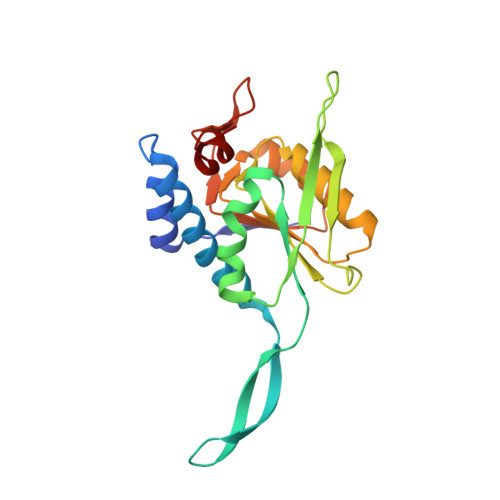
Uracil phosphoribosyltransferase (UPRTase) is part of the salvage pathway that leads to the biosynthesis of UMP. It catalyzes the formation of UMP and pyrophosphate from uracil and alpha-D-5-phosphoribosyl-1-pyrophosphate. Unlike enzymes in the de novo synthesis of UMP, UPRTases have only been found in lower organisms and are therefore potential targets for the development of new antibiotics. UPRTase from Bacillus caldolyticus has been crystallized and the structure has been determined by isomorphous replacement and refined to 3.0 A resolution. UPRTase from B. caldolyticus forms a dimer with the active sites pointing away from each other. A long arm from each subunit wraps around the other subunit, contributing half of the dimer interface. The monomer adopts the phosphoribosyltransferase type I fold, with a small C-terminal hood defining the uracil-binding site. The structure contains a well defined UMP molecule in the active site. The binding of UMP involves two sequence segments that are highly conserved among UPRTases. The first segment, Asp131-Ser139, contains the PRPP-binding consensus sequence motif known from other type I phosphoribosyltransferases and binds the ribose-5'-phosphate part of UMP. The second segment, Tyr193-Ala201, which is specific for uracil phosphoribosyltransferases, binds the uracil part of UMP through backbone contacts, partly mediated by a water molecule. Modelling of a PRPP-enzyme complex reveals that uracil can be activated to its tautomeric enol form by the complex. This is consistent with kinetic data, which display ordered sequential binding of substrates, with PRPP binding first. Based on this observation, a reaction mechanism is proposed.
Organizational Affiliation:
Centre for Crystallographic Studies, Department of Chemistry, University of Copenhagen, Universitetsparken 5, DK-2100 Copenhagen, Denmark. anders@ccs.ki.ku.dk