1I3Z

MURINE EAT2 SH2 DOMAIN IN COMPLEX WITH SLAM PHOSPHOPEPTIDE


Experimental Data Snapshot

  • Method: X-RAY DIFFRACTION
  • Resolution: 2.15 Å
  • R-Value Free: 0.279 
  • R-Value Work: 0.216 
  • R-Value Observed: 0.219 

wwPDB Validation   3D Report Full Report


This is version 1.3 of the entry. See complete history


Literature

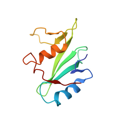
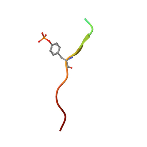
Structural basis for the interaction of the free SH2 domain EAT-2 with SLAM receptors in hematopoietic cells.

Morra, M.Lu, J.Poy, F.Martin, M.Sayos, J.Calpe, S.Gullo, C.Howie, D.Rietdijk, S.Thompson, A.Coyle, A.J.Denny, C.Yaffe, M.B.Engel, P.Eck, M.J.Terhorst, C.

(2001) Eur J Biochem 20: 5840-5852

  • DOI: https://doi.org/10.1093/emboj/20.21.5840
  • Primary Citation of Related Structures:  
    1I3Z

  • PubMed Abstract: 

    The T and natural killer (NK) cell-specific gene SAP (SH2D1A) encodes a 'free SH2 domain' that binds a specific tyrosine motif in the cytoplasmic tail of SLAM (CD150) and related cell surface proteins. Mutations in SH2D1A cause the X-linked lymphoproliferative disease, a primary immunodeficiency. Here we report that a second gene encoding a free SH2 domain, EAT-2, is expressed in macrophages and B lympho cytes. The EAT-2 structure in complex with a phosphotyrosine peptide containing a sequence motif with Tyr281 of the cytoplasmic tail of CD150 is very similar to the structure of SH2D1A complexed with the same peptide. This explains the high affinity of EAT-2 for the pTyr motif in the cytoplasmic tail of CD150 but, unlike SH2D1A, EAT-2 does not bind to non-phosphorylated CD150. EAT-2 binds to the phosphorylated receptors CD84, CD150, CD229 and CD244, and acts as a natural inhibitor, which interferes with the recruitment of the tyrosine phosphatase SHP-2. We conclude that EAT-2 plays a role in controlling signal transduction through at least four receptors expressed on the surface of professional antigen-presenting cells.


  • Organizational Affiliation

    Division of Immunology, Beth Israel Deaconess Medical Center, Harvard Medical School, Boston, MA 02115, USA. mmorra@caregroup.harvard.edu


Macromolecules
Find similar proteins by:  (by identity cutoff)  |  3D Structure
Entity ID: 1
MoleculeChains Sequence LengthOrganismDetailsImage
EWS/FLI1 ACTIVATED TRANSCRIPT 2103Mus musculusMutation(s): 0 
UniProt
Find proteins for O35324 (Mus musculus)
Explore O35324 
Go to UniProtKB:  O35324
Entity Groups  
Sequence Clusters30% Identity50% Identity70% Identity90% Identity95% Identity100% Identity
UniProt GroupO35324
Sequence Annotations
Expand
  • Reference Sequence

Find similar proteins by:  Sequence   |   3D Structure  

Entity ID: 2
MoleculeChains Sequence LengthOrganismDetailsImage
SIGNALING LYMPHOCYTIC ACTIVATION MOLECULE14N/AMutation(s): 1 
UniProt & NIH Common Fund Data Resources
Find proteins for Q13291 (Homo sapiens)
Explore Q13291 
Go to UniProtKB:  Q13291
PHAROS:  Q13291
GTEx:  ENSG00000117090 
Entity Groups  
Sequence Clusters30% Identity50% Identity70% Identity90% Identity95% Identity100% Identity
UniProt GroupQ13291
Sequence Annotations
Expand
  • Reference Sequence
Small Molecules
Modified Residues  1 Unique
IDChains TypeFormula2D DiagramParent
PTR
Query on PTR
B
L-PEPTIDE LINKINGC9 H12 N O6 PTYR
Experimental Data & Validation

Experimental Data

  • Method: X-RAY DIFFRACTION
  • Resolution: 2.15 Å
  • R-Value Free: 0.279 
  • R-Value Work: 0.216 
  • R-Value Observed: 0.219 
  • Space Group: P 21 21 2
Unit Cell:
Length ( Å )Angle ( ˚ )
a = 58.994α = 90
b = 59.5β = 90
c = 34.942γ = 90
Software Package:
Software NamePurpose
DENZOdata reduction
SCALEPACKdata scaling
AMoREphasing
REFMACrefinement

Structure Validation

View Full Validation Report


Entry History 

Deposition Data

Revision History  (Full details and data files)

  • Version 1.0: 2003-04-08
    Type: Initial release
  • Version 1.1: 2008-04-27
    Changes: Version format compliance
  • Version 1.2: 2011-07-13
    Changes: Version format compliance
  • Version 1.3: 2024-04-03
    Changes: Data collection, Database references, Derived calculations, Refinement description