1I2W

BETA-LACTAMASE FROM BACILLUS LICHENIFORMIS BS3 COMPLEXED WITH CEFOXITIN


Experimental Data Snapshot

  • Method: X-RAY DIFFRACTION
  • Resolution: 1.70 Å
  • R-Value Free: 0.256 
  • R-Value Work: 0.216 
  • R-Value Observed: 0.216 

wwPDB Validation   3D Report Full Report


Ligand Structure Quality Assessment 


This is version 2.0 of the entry. See complete history


Literature

Crystal structures of the Bacillus licheniformis BS3 class A beta-lactamase and of the acyl-enzyme adduct formed with cefoxitin

Fonze, E.Vanhove, M.Dive, G.Sauvage, E.Frere, J.M.Charlier, P.

(2002) Biochemistry 41: 1877-1885

  • DOI: https://doi.org/10.1021/bi015789k
  • Primary Citation of Related Structures:  
    1I2S, 1I2W

  • PubMed Abstract: 

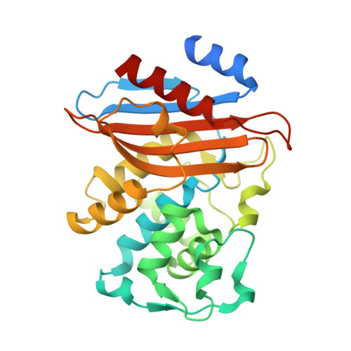
    The Bacillus licheniformis BS3 beta-lactamase catalyzes the hydrolysis of the beta-lactam ring of penicillins, cephalosporins, and related compounds. The production of beta-lactamases is the most common and thoroughly studied cause of antibiotic resistance. Although they escape the hydrolytic activity of the prototypical Staphylococcus aureus beta-lactamase, many cephems are good substrates for a large number of beta-lactamases. However, the introduction of a 7alpha-methoxy substituent, as in cefoxitin, extends their antibacterial spectrum to many cephalosporin-resistant Gram-negative bacteria. The 7alpha-methoxy group selectively reduces the hydrolytic action of many beta-lactamases without having a significant effect on the affinity for the target enzymes, the membrane penicillin-binding proteins. We report here the crystallographic structures of the BS3 enzyme and its acyl-enzyme adduct with cefoxitin at 1.7 A resolution. The comparison of the two structures reveals a covalent acyl-enzyme adduct with perturbed active site geometry, involving a different conformation of the omega-loop that bears the essential catalytic Glu166 residue. This deformation is induced by the cefoxitin side chain whose position is constrained by the presence of the alpha-methoxy group. The hydrolytic water molecule is also removed from the active site by the 7beta-carbonyl of the acyl intermediate. In light of the interactions and steric hindrances in the active site of the structure of the BS3-cefoxitin acyl-enzyme adduct, the crucial role of the conserved Asn132 residue is confirmed and a better understanding of the kinetic results emerges.


  • Organizational Affiliation

    Centre d'Ingénierie des Protéines, Institut de Physique B5 and Institut de Chimie B6, Université de Liège, B-4000 Sart Tilman, Belgium.


Macromolecules
Find similar proteins by:  (by identity cutoff)  |  3D Structure
Entity ID: 1
MoleculeChains Sequence LengthOrganismDetailsImage
BETA-LACTAMASE
A, B
282Bacillus licheniformisMutation(s): 0 
EC: 3.5.2.6
UniProt
Find proteins for P00808 (Bacillus licheniformis)
Explore P00808 
Go to UniProtKB:  P00808
Entity Groups  
Sequence Clusters30% Identity50% Identity70% Identity90% Identity95% Identity100% Identity
UniProt GroupP00808
Sequence Annotations
Expand
  • Reference Sequence
Small Molecules
Ligands 2 Unique
IDChains Name / Formula / InChI Key2D Diagram3D Interactions
1QL
Query on 1QL

Download Ideal Coordinates CCD File 
C [auth A],
D [auth B]
(2R)-5-[(carbamoyloxy)methyl]-2-{(1S)-1-methoxy-2-oxo-1-[(thiophen-2-ylacetyl)amino]ethyl}-3,6-dihydro-2H-1,3-thiazine-4-carboxylic acid
C16 H19 N3 O7 S2
IYZWSAWEHPQLHS-ZBFHGGJFSA-N
OUT
Query on OUT

Download Ideal Coordinates CCD File 
E [auth B]CARBAMIC ACID
C H3 N O2
KXDHJXZQYSOELW-UHFFFAOYSA-N
Experimental Data & Validation

Experimental Data

  • Method: X-RAY DIFFRACTION
  • Resolution: 1.70 Å
  • R-Value Free: 0.256 
  • R-Value Work: 0.216 
  • R-Value Observed: 0.216 
  • Space Group: P 1 21 1
Unit Cell:
Length ( Å )Angle ( ˚ )
a = 47.344α = 90
b = 106.372β = 94.18
c = 63.873γ = 90
Software Package:
Software NamePurpose
AMoREphasing
X-PLORrefinement
MAR345data collection
MOSFLMdata reduction
CCP4data scaling
Agrovatadata scaling
TRUNCATEdata scaling

Structure Validation

View Full Validation Report


Ligand Structure Quality Assessment 


Entry History 

Deposition Data

Revision History  (Full details and data files)

  • Version 1.0: 2002-03-13
    Type: Initial release
  • Version 1.1: 2008-04-27
    Changes: Version format compliance
  • Version 1.2: 2011-07-13
    Changes: Version format compliance
  • Version 1.3: 2017-10-04
    Changes: Refinement description
  • Version 2.0: 2018-09-12
    Changes: Advisory, Atomic model, Data collection, Derived calculations, Non-polymer description, Structure summary