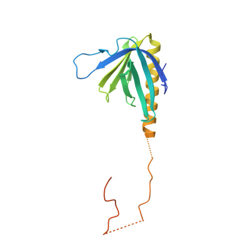
Crystal Structure of the Homer 1 Family Conserved Region Reveals the Interaction Between the EVH1 Domain and Own Proline-rich Motif
Irie, K., Nakatsu, T., Mitsuoka, K., Miyazawa, A., Sobue, K., Hiroaki, Y., Doi, T., Fujiyoshi, Y., Kato, H.(2002) J Mol Biol 318: 1117-1126
- PubMed: 12054806
- DOI: https://doi.org/10.1016/S0022-2836(02)00170-5
- Primary Citation of Related Structures:
1I2H - PubMed Abstract:
PSD-Zip45 (also named Homer 1c/Vesl-1L) is a synaptic scaffolding protein, which interacts with neurotransmitter receptors and other scaffolding proteins to target them into post-synaptic density (PSD), a specialized protein complex at the synaptic junction. Binding of the PSD-Zip45 to the receptors and scaffolding proteins results in colocalization and clustering of its binding partners in PSD. It has an Ena/VASP homology 1 (EVH1) domain in the N terminus for receptor binding, two leucine zipper motifs in the C terminus for clustering, and a linking region whose function is unclear despite the high level of conservation within the Homer 1 family. The X-ray crystallographic analysis of the largest fragment of residues 1-163, including an EVH1 domain reported here, demonstrates that the EVH1 domain contains an alpha-helix longer than that of the previous models, and that the linking part included in the conserved region of Homer 1 (CRH1) of the PSD-Zip45 interacts with the EVH1 domain of the neighbour CRH1 molecule in the crystal. The results suggest that the EVH1 domain recognizes the PPXXF motif found in the binding partners, and the SPLTP sequence (P-motif) in the linking region of the CRH1. The two types of binding are partly overlapped in the EVH1 domain, implying a mechanism to regulate multimerization of Homer 1 family proteins.
Organizational Affiliation:
Department of Biophysics, Kyoto University Graduate School of Science, Oiwake-cho, Kitashirakawa, Sakyo-ku, 606-8502, Japan.


