1HZO

STRUCTURE OF CLASS A CEPHALOSPORINASE FROM PROTEUS VULGARIS K1


Experimental Data Snapshot

  • Method: X-RAY DIFFRACTION
  • Resolution: 1.75 Å
  • R-Value Free: 0.193 
  • R-Value Work: 0.169 

wwPDB Validation   3D Report Full Report


This is version 1.4 of the entry. See complete history


Literature

Structure of an extended-spectrum class A beta-lactamase from Proteus vulgaris K1.

Nukaga, M.Mayama, K.Crichlow, G.V.Knox, J.R.

(2002) J Mol Biol 317: 109-117

  • DOI: https://doi.org/10.1006/jmbi.2002.5420
  • Primary Citation of Related Structures:  
    1HZO

  • PubMed Abstract: 

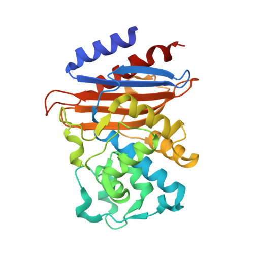
    The structure of a chromosomal extended-spectrum beta-lactamase (ESBL) having the ability to hydrolyze cephalosporins including cefuroxime and ceftazidime has been determined by X-ray crystallography to 1.75 A resolution. The species-specific class A beta-lactamase from Proteus vulgaris K1 was crystallized at pH 6.25 and its structure solved by molecular replacement. Refinement of the model resulted in crystallographic R and R(free) of 16.9 % and 19.3 %, respectively. The folding of the K1 enzyme is broadly similar to that of non-ESBL TEM-type beta-lactamases (2 A rmsd for C(alpha)) and differs by only 0.35 A for all atoms of six conserved residues in the catalytic site. Other residues promoting extended-spectrum activity in K1 include the side-chains of atypical residues Ser237 and Lys276. These side-chains are linked by two water molecules, one of which lies in the position normally filled by the guanidinium group of Arg244, present in most non-ESBL enzymes but absent from K1. The ammonium group of Lys276, ca 3.5 A from the virtual Arg244 guanidinium position, may interact with polar R2 substitutents on the dihydrothiazene ring of cephalosporins.


  • Organizational Affiliation

    Department of Molecular and Cell Biology, The University of Connecticut, Storrs, CT 06269-3125, USA.


Macromolecules
Find similar proteins by:  (by identity cutoff)  |  3D Structure
Entity ID: 1
MoleculeChains Sequence LengthOrganismDetailsImage
BETA-LACTAMASE
A, B
271Proteus vulgarisMutation(s): 0 
Gene Names: BLAC
EC: 3.5.2.6
UniProt
Find proteins for P52664 (Proteus vulgaris)
Explore P52664 
Go to UniProtKB:  P52664
Entity Groups  
Sequence Clusters30% Identity50% Identity70% Identity90% Identity95% Identity100% Identity
UniProt GroupP52664
Sequence Annotations
Expand
  • Reference Sequence
Small Molecules
Ligands 1 Unique
IDChains Name / Formula / InChI Key2D Diagram3D Interactions
MES
Query on MES

Download Ideal Coordinates CCD File 
C [auth A],
D [auth B]
2-(N-MORPHOLINO)-ETHANESULFONIC ACID
C6 H13 N O4 S
SXGZJKUKBWWHRA-UHFFFAOYSA-N
Experimental Data & Validation

Experimental Data

  • Method: X-RAY DIFFRACTION
  • Resolution: 1.75 Å
  • R-Value Free: 0.193 
  • R-Value Work: 0.169 
  • Space Group: P 21 21 21
Unit Cell:
Length ( Å )Angle ( ˚ )
a = 58.65α = 90
b = 66.15β = 90
c = 134.394γ = 90
Software Package:
Software NamePurpose
AMoREphasing
CNSrefinement
X-GENdata reduction
X-GENdata scaling

Structure Validation

View Full Validation Report


Entry History 

Deposition Data

Revision History  (Full details and data files)

  • Version 1.0: 2002-04-03
    Type: Initial release
  • Version 1.1: 2008-04-27
    Changes: Version format compliance
  • Version 1.2: 2011-07-13
    Changes: Version format compliance
  • Version 1.3: 2017-10-04
    Changes: Refinement description
  • Version 1.4: 2023-08-09
    Changes: Data collection, Database references, Derived calculations, Refinement description