1HZJ

HUMAN UDP-GALACTOSE 4-EPIMERASE: ACCOMMODATION OF UDP-N-ACETYLGLUCOSAMINE WITHIN THE ACTIVE SITE


Experimental Data Snapshot

  • Method: X-RAY DIFFRACTION
  • Resolution: 1.50 Å
  • R-Value Free: 0.227 
  • R-Value Work: 0.184 
  • R-Value Observed: 0.185 

wwPDB Validation   3D Report Full Report


Ligand Structure Quality Assessment 


This is version 1.3 of the entry. See complete history


Literature

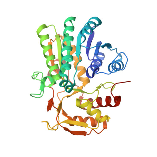
Human UDP-galactose 4-epimerase. Accommodation of UDP-N-acetylglucosamine within the active site.

Thoden, J.B.Wohlers, T.M.Fridovich-Keil, J.L.Holden, H.M.

(2001) J Biol Chem 276: 15131-15136

  • DOI: https://doi.org/10.1074/jbc.M100220200
  • Primary Citation of Related Structures:  
    1HZJ

  • PubMed Abstract: 

    UDP-galactose 4-epimerase catalyzes the interconversion of UDP-galactose and UDP-glucose during normal galactose metabolism. One of the key structural features in the proposed reaction mechanism for the enzyme is the rotation of a 4'-ketopyranose intermediate within the active site pocket. Recently, the three-dimensional structure of the human enzyme with bound NADH and UDP-glucose was determined. Unlike that observed for the protein isolated from Escherichia coli, the human enzyme can also turn over UDP-GlcNAc to UDP-GalNAc and vice versa. Here we describe the three-dimensional structure of human epimerase complexed with NADH and UDP-GlcNAc. To accommodate the additional N-acetyl group at the C2 position of the sugar, the side chain of Asn-207 rotates toward the interior of the protein and interacts with Glu-199. Strikingly, in the human enzyme, the structural equivalent of Tyr-299 in the E. coli protein is replaced with a cysteine residue (Cys-307) and the active site volume for the human protein is calculated to be approximately 15% larger than that observed for the bacterial epimerase. This combination of a larger active site cavity and amino acid residue replacement most likely accounts for the inability of the E. coli enzyme to interconvert UDP-GlcNAc and UDP-GalNAc.


  • Organizational Affiliation

    Department of Biochemistry, University of Wisconsin, Madison, Wisconsin 53705, USA.


Macromolecules
Find similar proteins by:  (by identity cutoff)  |  3D Structure
Entity ID: 1
MoleculeChains Sequence LengthOrganismDetailsImage
UDP-GALACTOSE 4-EPIMERASE
A, B
348Homo sapiensMutation(s): 0 
EC: 5.1.3.2
UniProt & NIH Common Fund Data Resources
Find proteins for Q14376 (Homo sapiens)
Explore Q14376 
Go to UniProtKB:  Q14376
PHAROS:  Q14376
GTEx:  ENSG00000117308 
Entity Groups  
Sequence Clusters30% Identity50% Identity70% Identity90% Identity95% Identity100% Identity
UniProt GroupQ14376
Sequence Annotations
Expand
  • Reference Sequence
Small Molecules
Ligands 4 Unique
IDChains Name / Formula / InChI Key2D Diagram3D Interactions
NAD
Query on NAD

Download Ideal Coordinates CCD File 
F [auth A],
I [auth B]
NICOTINAMIDE-ADENINE-DINUCLEOTIDE
C21 H27 N7 O14 P2
BAWFJGJZGIEFAR-NNYOXOHSSA-N
UD1
Query on UD1

Download Ideal Coordinates CCD File 
G [auth A],
J [auth B]
URIDINE-DIPHOSPHATE-N-ACETYLGLUCOSAMINE
C17 H27 N3 O17 P2
LFTYTUAZOPRMMI-CFRASDGPSA-N
CL
Query on CL

Download Ideal Coordinates CCD File 
C [auth A],
D [auth A],
H [auth B]
CHLORIDE ION
Cl
VEXZGXHMUGYJMC-UHFFFAOYSA-M
MG
Query on MG

Download Ideal Coordinates CCD File 
E [auth A]MAGNESIUM ION
Mg
JLVVSXFLKOJNIY-UHFFFAOYSA-N
Experimental Data & Validation

Experimental Data

  • Method: X-RAY DIFFRACTION
  • Resolution: 1.50 Å
  • R-Value Free: 0.227 
  • R-Value Work: 0.184 
  • R-Value Observed: 0.185 
  • Space Group: P 21 21 21
Unit Cell:
Length ( Å )Angle ( ˚ )
a = 63.4α = 90
b = 88.9β = 90
c = 118.7γ = 90
Software Package:
Software NamePurpose
TNTrefinement
d*TREKdata reduction
SCALEPACKdata scaling
TNTphasing

Structure Validation

View Full Validation Report


Ligand Structure Quality Assessment 


Entry History 

Deposition Data

Revision History  (Full details and data files)

  • Version 1.0: 2001-05-09
    Type: Initial release
  • Version 1.1: 2008-04-27
    Changes: Version format compliance
  • Version 1.2: 2011-07-13
    Changes: Version format compliance
  • Version 1.3: 2023-08-09
    Changes: Data collection, Database references, Derived calculations, Refinement description