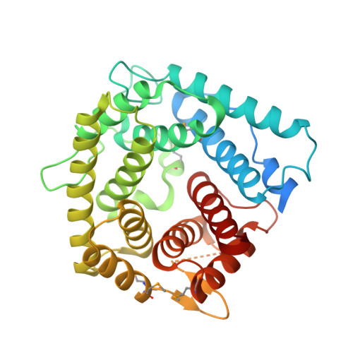
X-ray crystal structure of the C4d fragment of human complement component C4.
van den Elsen, J.M., Martin, A., Wong, V., Clemenza, L., Rose, D.R., Isenman, D.E.(2002) J Mol Biol 322: 1103-1115
- PubMed: 12367531
- DOI: https://doi.org/10.1016/s0022-2836(02)00854-9
- Primary Citation of Related Structures:
1HZF - PubMed Abstract:
C4 fulfills a vital role in the propagation of the classical and lectin pathways of the complement system. Although there are no reports to date of a C4 functional activity that is mediated solely by the C4d region, evidence clearly points to it having a vital role in a number of the properties of native C4 and its major activation fragment, C4b. Contained within the C4d region are the thioester-forming residues, the four isotype-specific residues controlling the C4A/C4B transacylation preferences, a binding site for nascent C3b important in assembling the classical pathway C5 convertase and determinants for the Chido/Rodgers (Ch/Rg) blood group antigens. In view of its functional importance, we undertook to determine the three-dimensional structure of C4d by X-ray crystallography. Here we report the 2.3A resolution structure of C4Ad, the C4d fragment derived from the human C4A isotype. Although the approximately 30% sequence identity between C4Ad and the corresponding fragment of C3 might be expected to establish a general fold similarity between the two molecules, C4Ad in fact displays a fold that is essentially superimposable on the structure of C3d. By contrast, the electrostatic characteristics of the various faces of the C4Ad molecule show marked differences from the corresponding faces of C3d, likely reflecting the differences in function between C3 and C4. Residues previously predicted to form the major Ch/Rg epitopes were proximately located and accessible on the concave surface of C4Ad. In addition to providing further insights on the current models for the covalent binding reaction, the C4Ad structure allows one to rationalize why C4d is not a ligand for complement receptor 2. Finally the structure allows for the visualization of the face of the molecule containing the binding site for C3b utilized in the assembly of classical pathway C5 convertase.
Organizational Affiliation:
Ontario Cancer Institute and Department of Medical Biophysics, University of Toronto, Ontario, Canada.