Structures of the B1 domain of protein L from Peptostreptococcus magnus with a tyrosine to tryptophan substitution.
O'Neill, J.W., Kim, D.E., Baker, D., Zhang, K.Y.(2001) Acta Crystallogr D Biol Crystallogr 57: 480-487
- PubMed: 11264576
- DOI: https://doi.org/10.1107/s0907444901000373
- Primary Citation of Related Structures:
1HZ5, 1HZ6 - PubMed Abstract:
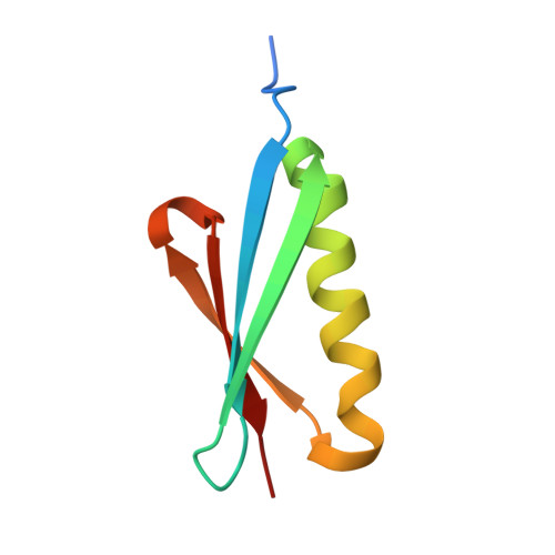
The three-dimensional structure of a tryptophan-containing variant of the IgG-binding B1 domain of protein L has been solved in two crystal forms to 1.7 and 1.8 A resolution. In one of the crystal forms, the entire N-terminal histidine-tag region was immobilized through the coordination of zinc ions and its structural conformation along with the zinc coordination scheme were determined. However, the ordering of the histidine tag by zinc does not affect the overall structure of the rest of the protein. Structural comparisons of the tryptophan-containing variant with an NMR-derived wild-type structure, which contains a tyrosine at position 47, reveals a common fold, although the overall backbone root-mean-square difference is 1.5 A. The Y47W substitution only caused local rearrangement of several side chains, the most prominent of which is the rotation of the Tyr34 side chain, resulting in a 6 A displacement of its hydroxyl group. A small methyl-sized cavity bounded by beta-strands 1, 2 and 4 and the alpha-helix was found in the structures of the Y47W-substituted protein L B1 domain. This cavity may be created as the result of subsequent side-chain rearrangements caused by the Y47W substitution. These high-resolution structures of the tryptophan-containing variant provide a reference frame for the analysis of thermodynamic and kinetic data derived from a series of mutational studies of the protein L B1 domain.
Organizational Affiliation:
Division of Basic Sciences, Fred Hutchinson Cancer Research Center, 1100 Fairview Avenue North, Seattle, WA 98109, USA.