1HYO

CRYSTAL STRUCTURE OF FUMARYLACETOACETATE HYDROLASE COMPLEXED WITH 4-(HYDROXYMETHYLPHOSPHINOYL)-3-OXO-BUTANOIC ACID


Experimental Data Snapshot

  • Method: X-RAY DIFFRACTION
  • Resolution: 1.30 Å
  • R-Value Free: 0.199 
  • R-Value Work: 0.181 
  • R-Value Observed: 0.181 

wwPDB Validation   3D Report Full Report


Ligand Structure Quality Assessment 


This is version 1.3 of the entry. See complete history


Literature

Mechanistic inferences from the crystal structure of fumarylacetoacetate hydrolase with a bound phosphorus-based inhibitor.

Bateman, R.L.Bhanumoorthy, P.Witte, J.F.McClard, R.W.Grompe, M.Timm, D.E.

(2001) J Biol Chem 276: 15284-15291

  • DOI: https://doi.org/10.1074/jbc.M007621200
  • Primary Citation of Related Structures:  
    1HYO

  • PubMed Abstract: 

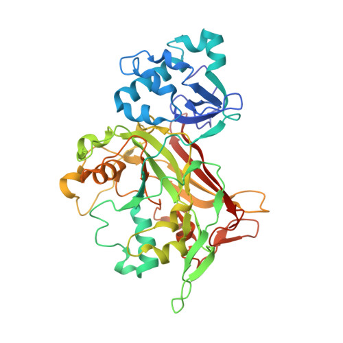
    Fumarylacetoacetate hydrolase (FAH) catalyzes the hydrolytic cleavage of a carbon-carbon bond in fumarylacetoacetate to yield fumarate and acetoacetate as the final step of Phe and Tyr degradation. This unusual reaction is an essential human metabolic function, with loss of FAH activity causing the fatal metabolic disease hereditary tyrosinemia type I (HT1). An enzymatic mechanism involving a catalytic metal ion, a Glu/His catalytic dyad, and a charged oxyanion hole was previously proposed based on recently determined FAH crystal structures. Here we report the development and characterization of an FAH inhibitor, 4-(hydroxymethylphosphinoyl)-3-oxo-butanoic acid (HMPOBA), that competes with the physiological substrate with a K(i) of 85 microM. The crystal structure of FAH complexed with HMPOBA refined at 1.3-A resolution reveals the molecular basis for the competitive inhibition, supports the proposed formation of a tetrahedral alkoxy transition state intermediate during the FAH catalyzed reaction, and reveals a Mg(2+) bound in the enzyme's active site. The analysis of FAH structures corresponding to different catalytic states reveals significant active site side-chain motions that may also be related to catalytic function. Thus, these results advance the understanding of an essential catabolic reaction associated with a fatal metabolic disease and provide insight into the structure-based development of FAH inhibitors.


  • Organizational Affiliation

    Department of Molecular and Medical Genetics, Oregon Health Sciences University, and the Department of Chemistry, Reed College, Portland, Oregon, USA.


Macromolecules
Find similar proteins by:  (by identity cutoff)  |  3D Structure
Entity ID: 1
MoleculeChains Sequence LengthOrganismDetailsImage
FUMARYLACETOACETATE HYDROLASE
A, B
421Mus musculusMutation(s): 0 
EC: 3.7.1.2
UniProt & NIH Common Fund Data Resources
Find proteins for P35505 (Mus musculus)
Explore P35505 
Go to UniProtKB:  P35505
IMPC:  MGI:95482
Entity Groups  
Sequence Clusters30% Identity50% Identity70% Identity90% Identity95% Identity100% Identity
UniProt GroupP35505
Sequence Annotations
Expand
  • Reference Sequence
Small Molecules
Ligands 5 Unique
IDChains Name / Formula / InChI Key2D Diagram3D Interactions
HBU
Query on HBU

Download Ideal Coordinates CCD File 
F [auth A],
K [auth B]
4-[HYDROXY-[METHYL-PHOSPHINOYL]]-3-OXO-BUTANOIC ACID
C5 H9 O5 P
MZTALSPOBPYJPG-UHFFFAOYSA-N
ACT
Query on ACT

Download Ideal Coordinates CCD File 
E [auth A],
J [auth B]
ACETATE ION
C2 H3 O2
QTBSBXVTEAMEQO-UHFFFAOYSA-M
NI
Query on NI

Download Ideal Coordinates CCD File 
I [auth B]NICKEL (II) ION
Ni
VEQPNABPJHWNSG-UHFFFAOYSA-N
CA
Query on CA

Download Ideal Coordinates CCD File 
D [auth A],
H [auth B]
CALCIUM ION
Ca
BHPQYMZQTOCNFJ-UHFFFAOYSA-N
MG
Query on MG

Download Ideal Coordinates CCD File 
C [auth A],
G [auth B]
MAGNESIUM ION
Mg
JLVVSXFLKOJNIY-UHFFFAOYSA-N
Binding Affinity Annotations 
IDSourceBinding Affinity
HBU Binding MOAD:  1HYO Ki: 8.48e+4 (nM) from 1 assay(s)
PDBBind:  1HYO Ki: 8.48e+4 (nM) from 1 assay(s)
Experimental Data & Validation

Experimental Data

  • Method: X-RAY DIFFRACTION
  • Resolution: 1.30 Å
  • R-Value Free: 0.199 
  • R-Value Work: 0.181 
  • R-Value Observed: 0.181 
  • Space Group: P 1 21 1
Unit Cell:
Length ( Å )Angle ( ˚ )
a = 64.102α = 90
b = 109.458β = 102.37
c = 67.459γ = 90
Software Package:
Software NamePurpose
AMoREphasing
REFMACrefinement
DENZOdata reduction
SCALEPACKdata scaling

Structure Validation

View Full Validation Report


Ligand Structure Quality Assessment 


Entry History 

Deposition Data

Revision History  (Full details and data files)

  • Version 1.0: 2001-02-14
    Type: Initial release
  • Version 1.1: 2008-04-27
    Changes: Version format compliance
  • Version 1.2: 2011-07-13
    Changes: Version format compliance
  • Version 1.3: 2023-08-09
    Changes: Data collection, Database references, Derived calculations, Refinement description