1HWT

STRUCTURE OF A HAP1/DNA COMPLEX REVEALS DRAMATICALLY ASYMMETRIC DNA BINDING BY A HOMODIMERIC PROTEIN


Experimental Data Snapshot

  • Method: X-RAY DIFFRACTION
  • Resolution: 2.50 Å
  • R-Value Free: 0.291 
  • R-Value Work: 0.246 
  • R-Value Observed: 0.246 

wwPDB Validation   3D Report Full Report


This is version 1.4 of the entry. See complete history


Literature

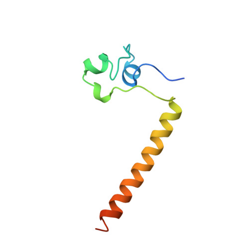
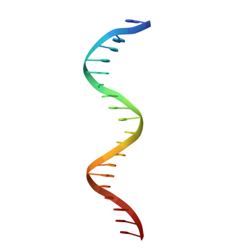
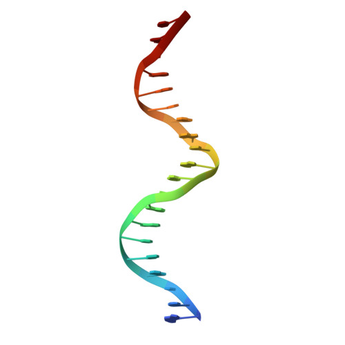
Structure of a HAP1-DNA complex reveals dramatically asymmetric DNA binding by a homodimeric protein.

King, D.A.Zhang, L.Guarente, L.Marmorstein, R.

(1999) Nat Struct Biol 6: 64-71

  • DOI: https://doi.org/10.1038/4940
  • Primary Citation of Related Structures:  
    1HWT

  • PubMed Abstract: 

    HAP1 is a member of a family of fungal transcription factors that contain a Zn2Cys6 binuclear cluster domain and bind as homodimers to sequences containing two DNA half sites. We have determined the 2.5 A crystal structure of HAP1 bound to a cognate upstream activation sequence from the CYC7 gene. The structure reveals that HAP1 is bound in a dramatically asymmetric manner to the DNA target. This asymmetry aligns the Zn2Cys6 domains in a tandem head-to-tail fashion to contact two DNA half sites, positions an N-terminal arm of one of the protein subunits to interact with the inter-half site base pairs in the DNA minor groove, and suggests a mechanism by which DNA-binding facilitates asymmetric dimerization by HAP1. Comparisons with the DNA complexes of the related GAL4, PPR1 and PUT3 proteins illustrate how a conserved protein domain can be reoriented to recognize DNA half sites of different polarities and how homodimeric proteins adopt dramatically asymmetric structures to recognize cognate DNA targets.


  • Organizational Affiliation

    The Wistar Institute and The Department of Chemistry, University of Pennsylvania, Philadelphia 19104, USA.


Macromolecules

Find similar proteins by:  (by identity cutoff)  |  3D Structure
Entity ID: 3
MoleculeChains Sequence LengthOrganismDetailsImage
PROTEIN (HEME ACTIVATOR PROTEIN)E [auth C],
F [auth D],
G,
H
81Saccharomyces cerevisiaeMutation(s): 0 
UniProt
Find proteins for P0CS82 (Saccharomyces cerevisiae)
Explore P0CS82 
Go to UniProtKB:  P0CS82
Entity Groups  
Sequence Clusters30% Identity50% Identity70% Identity90% Identity95% Identity100% Identity
UniProt GroupP0CS82
Sequence Annotations
Expand
  • Reference Sequence

Find similar nucleic acids by:  Sequence   |   3D Structure  

Entity ID: 1
MoleculeChains LengthOrganismImage
DNA (5'-D(*GP*CP*GP*CP*TP*AP*TP*TP*AP*TP*CP*GP*CP*TP*AP*TP*TP*AP*GP*C)-3')A,
C [auth E]
20N/A
Sequence Annotations
Expand
  • Reference Sequence

Find similar nucleic acids by:  Sequence   |   3D Structure  

Entity ID: 2
MoleculeChains LengthOrganismImage
DNA (5'-D(*GP*CP*TP*AP*AP*TP*AP*GP*CP*GP*AP*TP*AP*AP*TP*AP*GP*CP*GP*C)-3')B,
D [auth F]
20N/A
Sequence Annotations
Expand
  • Reference Sequence
Experimental Data & Validation

Experimental Data

  • Method: X-RAY DIFFRACTION
  • Resolution: 2.50 Å
  • R-Value Free: 0.291 
  • R-Value Work: 0.246 
  • R-Value Observed: 0.246 
  • Space Group: P 21 21 21
Unit Cell:
Length ( Å )Angle ( ˚ )
a = 85.6α = 90
b = 85.7β = 90
c = 94γ = 90
Software Package:
Software NamePurpose
AMoREphasing
X-PLORrefinement
XDSdata reduction
XSCALEdata scaling

Structure Validation

View Full Validation Report


Entry History 

Deposition Data

Revision History  (Full details and data files)

  • Version 1.0: 1999-11-10
    Type: Initial release
  • Version 1.1: 2008-05-22
    Changes: Version format compliance
  • Version 1.2: 2011-07-13
    Changes: Version format compliance
  • Version 1.3: 2024-02-07
    Changes: Data collection, Database references, Derived calculations
  • Version 1.4: 2024-04-03
    Changes: Refinement description