1HUX

CRYSTAL STRUCTURE OF THE ACIDAMINOCOCCUS FERMENTANS (R)-2-HYDROXYGLUTARYL-COA DEHYDRATASE COMPONENT A


Experimental Data Snapshot

  • Method: X-RAY DIFFRACTION
  • Resolution: 3.00 Å
  • R-Value Free: 0.279 
  • R-Value Work: 0.236 

wwPDB Validation   3D Report Full Report


This is version 1.3 of the entry. See complete history


Literature

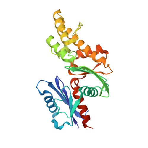
Crystal structure of the Acidaminococcus fermentans 2-hydroxyglutaryl-CoA dehydratase component A.

Locher, K.P.Hans, M.Yeh, A.P.Schmid, B.Buckel, W.Rees, D.C.

(2001) J Mol Biol 307: 297-308

  • DOI: https://doi.org/10.1006/jmbi.2000.4496
  • Primary Citation of Related Structures:  
    1HUX

  • PubMed Abstract: 

    Acidaminococcus fermentans degrades glutamate via the hydroxyglutarate pathway, which involves the syn-elimination of water from (R)-2-hydroxyglutaryl-CoA in a key reaction of the pathway. This anaerobic process is catalyzed by 2-hydroxyglutaryl-CoA dehydratase, an enzyme with two components (A and D) that reversibly associate during reaction cycles. Component A (CompA), a homodimeric protein of 2x27 kDa, contains a single, bridging [4Fe-4S] cluster and uses the hydrolysis of ATP to deliver an electron to the dehydratase component (CompD), where the electron is used catalytically. The structure of the extremely oxygen-sensitive CompA protein was solved by X-ray crystallography to 3 A resolution. The protein was found to be a member of the actin fold family, revealing a similar architecture and nucleotide-binding site. The key differences between CompA and other members of the actin fold family are: (i) the presence of a cluster binding segment, the "cluster helix"; (ii) the [4Fe-4S] cluster; and (iii) the location of the homodimer interface, which involves the bridging cluster. Possible reaction mechanisms are discussed in light of the close structural similarity to members of the actin-fold family and the functional similarity to the nitrogenase Fe- protein.


  • Organizational Affiliation

    Howard Hughes Medical Institute, Division of Chemistry and Chemical Engineering, California Institute of Technology, Mail Code 147-75CH, Pasadena, CA 91125, USA.


Macromolecules
Find similar proteins by:  (by identity cutoff)  |  3D Structure
Entity ID: 1
MoleculeChains Sequence LengthOrganismDetailsImage
ACTIVATOR OF (R)-2-HYDROXYGLUTARYL-COA DEHYDRATASE
A, B
270Acidaminococcus fermentansMutation(s): 0 
Gene Names: HGDC
UniProt
Find proteins for P11568 (Acidaminococcus fermentans (strain ATCC 25085 / DSM 20731 / CCUG 9996 / CIP 106432 / VR4))
Explore P11568 
Go to UniProtKB:  P11568
Entity Groups  
Sequence Clusters30% Identity50% Identity70% Identity90% Identity95% Identity100% Identity
UniProt GroupP11568
Sequence Annotations
Expand
  • Reference Sequence
Small Molecules
Ligands 2 Unique
IDChains Name / Formula / InChI Key2D Diagram3D Interactions
ADP
Query on ADP

Download Ideal Coordinates CCD File 
D [auth A],
E [auth B]
ADENOSINE-5'-DIPHOSPHATE
C10 H15 N5 O10 P2
XTWYTFMLZFPYCI-KQYNXXCUSA-N
SF4
Query on SF4

Download Ideal Coordinates CCD File 
C [auth A]IRON/SULFUR CLUSTER
Fe4 S4
LJBDFODJNLIPKO-UHFFFAOYSA-N
Experimental Data & Validation

Experimental Data

  • Method: X-RAY DIFFRACTION
  • Resolution: 3.00 Å
  • R-Value Free: 0.279 
  • R-Value Work: 0.236 
  • Space Group: P 1 21 1
Unit Cell:
Length ( Å )Angle ( ˚ )
a = 82.07α = 90
b = 51.99β = 118.81
c = 74.7γ = 90
Software Package:
Software NamePurpose
SHARPphasing
CNSrefinement
DENZOdata reduction
SCALEPACKdata scaling

Structure Validation

View Full Validation Report


Entry History 

Deposition Data

Revision History  (Full details and data files)

  • Version 1.0: 2001-03-21
    Type: Initial release
  • Version 1.1: 2008-04-27
    Changes: Version format compliance
  • Version 1.2: 2011-07-13
    Changes: Version format compliance
  • Version 1.3: 2024-02-07
    Changes: Data collection, Database references, Derived calculations