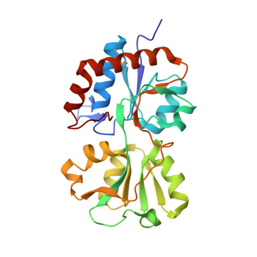
Refined 1.89-A structure of the histidine-binding protein complexed with histidine and its relationship with many other active transport/chemosensory proteins.
Yao, N., Trakhanov, S., Quiocho, F.A.(1994) Biochemistry 33: 4769-4779
- PubMed: 8161536
- DOI: https://doi.org/10.1021/bi00182a004
- Primary Citation of Related Structures:
1HSL - PubMed Abstract:
The structure of the histidine-binding protein (HBP, M(r) = 26,100), involved solely in active transport, has been determined by the molecular replacement technique and refined to 1.89-A resolution and to an R-factor of 0.199. The structure is that of two protein molecules, each with a bound L-histidine, in the asymmetric unit. Replacement solution was achieved by using a model of the crystal structure of the ligand-free, open-cleft form of the lysine/arginine/ornithine-binding protein which was modified so that the two domains are close to each other by bending the hinge connecting the two domains. The bound histidine is held in place by 10 hydrogen bonds, 2 salt links, and about 60 van der Waals contacts. Elucidation of the HBP structure brings a total of eight different binding proteins structures determined in our laboratory, including those with specificities for monosaccharides, maltodextrins (linear and cyclic), aliphatic amino acids, and inorganic oxyanions. These structures comprise about a third of the entire family of periplasmic binding proteins which act as initial primary high-affinity receptors of active transport in Gram-negative bacteria. Two of the binding proteins with specificities for glucose/galactose and maltodextrins also serve in a similar capacity in chemotaxis. Though these proteins have different molecular weights (ranging from 26,000 to 40,000), amino acid sequences, and ligand specificities, their three-dimensional structures are similar overall. They are elongated (axial ratios of 2:1) and composed of two similar globular domains separated by a deep cleft wherein the ligand-binding site is located. These structures provide understanding of molecular recognition of a variety of ligands at the atomic level and functional roles of the binding proteins.
Organizational Affiliation:
Department of Biochemistry, Howard Hughes Medical Institute, Houston, Texas 77030.