1HS6

STRUCTURE OF LEUKOTRIENE A4 HYDROLASE COMPLEXED WITH BESTATIN.


Experimental Data Snapshot

  • Method: X-RAY DIFFRACTION
  • Resolution: 1.95 Å
  • R-Value Free: 0.247 
  • R-Value Work: 0.188 
  • R-Value Observed: 0.185 

wwPDB Validation   3D Report Full Report


This is version 1.4 of the entry. See complete history


Literature

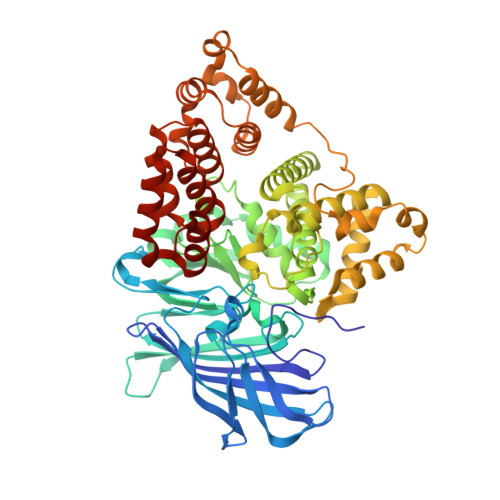
Crystal structure of human leukotriene A(4) hydrolase, a bifunctional enzyme in inflammation.

Thunnissen, M.M.Nordlund, P.Haeggstrom, J.Z.

(2001) Nat Struct Biol 8: 131-135

  • DOI: https://doi.org/10.1038/84117
  • Primary Citation of Related Structures:  
    1HS6

  • PubMed Abstract: 

    Leukotriene (LT) A(4) hydrolase/aminopeptidase (LTA4H) is a bifunctional zinc enzyme that catalyzes the biosynthesis of LTB4, a potent lipid chemoattractant involved in inflammation, immune responses, host defense against infection, and PAF-induced shock. The high resolution crystal structure of LTA4H in complex with the competitive inhibitor bestatin reveals a protein folded into three domains that together create a deep cleft harboring the catalytic Zn(2+) site. A bent and narrow pocket, shaped to accommodate the substrate LTA(4), constitutes a highly confined binding region that can be targeted in the design of specific anti-inflammatory agents. Moreover, the structure of the catalytic domain is very similar to that of thermolysin and provides detailed insight into mechanisms of catalysis, in particular the chemical strategy for the unique epoxide hydrolase reaction that generates LTB(4).


  • Organizational Affiliation

    Department of Biochemistry, University of Stockholm, Arrhenius Laboratories A4, S-106 91 Stockholm, Sweden. marjo@dbb.su.se


Macromolecules
Find similar proteins by:  (by identity cutoff)  |  3D Structure
Entity ID: 1
MoleculeChains Sequence LengthOrganismDetailsImage
LEUKOTRIENE A-4 HYDROLASE611Homo sapiensMutation(s): 0 
EC: 3.3.2.6
UniProt & NIH Common Fund Data Resources
Find proteins for P09960 (Homo sapiens)
Explore P09960 
Go to UniProtKB:  P09960
PHAROS:  P09960
GTEx:  ENSG00000111144 
Entity Groups  
Sequence Clusters30% Identity50% Identity70% Identity90% Identity95% Identity100% Identity
UniProt GroupP09960
Sequence Annotations
Expand
  • Reference Sequence
Small Molecules
Binding Affinity Annotations 
IDSourceBinding Affinity
BES BindingDB:  1HS6 Ki: 320 (nM) from 1 assay(s)
Kd: min: 2050, max: 5150 (nM) from 3 assay(s)
IC50: min: 266, max: 2.00e+4 (nM) from 8 assay(s)
PDBBind:  1HS6 IC50: 4200 (nM) from 1 assay(s)
Experimental Data & Validation

Experimental Data

  • Method: X-RAY DIFFRACTION
  • Resolution: 1.95 Å
  • R-Value Free: 0.247 
  • R-Value Work: 0.188 
  • R-Value Observed: 0.185 
  • Space Group: P 21 21 2
Unit Cell:
Length ( Å )Angle ( ˚ )
a = 67.6α = 90
b = 133.5β = 90
c = 83.4γ = 90
Software Package:
Software NamePurpose
MLPHAREphasing
SHARPphasing
TNTrefinement
DENZOdata reduction
SCALEPACKdata scaling

Structure Validation

View Full Validation Report


Entry History 

Deposition Data

Revision History  (Full details and data files)

  • Version 1.0: 2001-06-24
    Type: Initial release
  • Version 1.1: 2008-04-27
    Changes: Version format compliance
  • Version 1.2: 2011-07-13
    Changes: Version format compliance
  • Version 1.3: 2019-11-20
    Changes: Derived calculations
  • Version 1.4: 2024-03-13
    Changes: Data collection, Database references, Derived calculations