Subunit-subunit interactions in trimeric arginase. Generation of active monomers by mutation of a single amino acid.
Lavulo, L.T., Sossong Jr., T.M., Brigham-Burke, M.R., Doyle, M.L., Cox, J.D., Christianson, D.W., Ash, D.E.(2001) J Biol Chem 276: 14242-14248
- PubMed: 11278703
- DOI: https://doi.org/10.1074/jbc.M010575200
- Primary Citation of Related Structures:
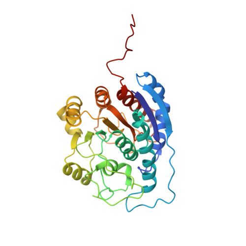
1HQX - PubMed Abstract:
The structure of the trimeric, manganese metalloenzyme, rat liver arginase, has been previously determined at 2.1-A resolution (Kanyo, Z. F., Scolnick, L. R., Ash, D. E., and Christianson, D. W., (1996) Nature 383, 554-557). A key feature of this structure is a novel S-shaped oligomerization motif at the carboxyl terminus of the protein that mediates approximately 54% of the intermonomer contacts. Arg-308, located within this oligomerization motif, nucleates a series of intramonomer and intermonomer salt links. In contrast to the trimeric wild-type enzyme, the R308A, R308E, and R308K variants of arginase exist as monomeric species, as determined by gel filtration and analytical ultracentrifugation, indicating that mutation of Arg-308 shifts the equilibrium for trimer dissociation by at least a factor of 10(5). These monomeric arginase variants are catalytically active, with k(cat)/K(m) values that are 13-17% of the value for wild-type enzyme. The arginase variants are characterized by decreased temperature stability relative to the wild-type enzyme. Differential scanning calorimetry shows that the midpoint temperature for unfolding of the Arg-308 variants is in the range of 63.6-65.5 degrees C, while the corresponding value for the wild-type enzyme is 70 degrees C. The three-dimensional structure of the R308K variant has been determined at 3-A resolution. At the high protein concentrations utilized in the crystallizations, this variant exists as a trimer, but weakened salt link interactions are observed for Lys-308.
Organizational Affiliation:
Department of Biochemistry, Temple University School of Medicine, Philadelphia, Pennsylvania 19140, USA.