1HKC

RECOMBINANT HUMAN HEXOKINASE TYPE I COMPLEXED WITH GLUCOSE AND PHOSPHATE


Experimental Data Snapshot

  • Method: X-RAY DIFFRACTION
  • Resolution: 2.80 Å
  • R-Value Free: 0.241 
  • R-Value Work: 0.174 
  • R-Value Observed: 0.174 

wwPDB Validation   3D Report Full Report


Ligand Structure Quality Assessment 


This is version 1.4 of the entry. See complete history


Literature

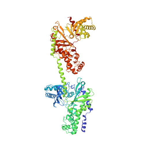
Regulation of hexokinase I: crystal structure of recombinant human brain hexokinase complexed with glucose and phosphate.

Aleshin, A.E.Zeng, C.Bartunik, H.D.Fromm, H.J.Honzatko, R.B.

(1998) J Mol Biol 282: 345-357

  • DOI: https://doi.org/10.1006/jmbi.1998.2017
  • Primary Citation of Related Structures:  
    1HKC

  • PubMed Abstract: 

    Hexokinase I, the pacemaker of glycolysis in brain tissue and red blood cells, is comprised of two similar domains fused into a single polypeptide chain. The C-terminal half of hexokinase I is catalytically active, whereas the N-terminal half is necessary for the relief of product inhibition by phosphate. A crystalline complex of recombinant human hexokinase I with glucose and phosphate (2.8 A resolution) reveals a single binding site for phosphate and glucose at the N-terminal half of the enzyme. Glucose and phosphate stabilize the N-terminal half in a closed conformation. Unexpectedly, glucose binds weakly to the C-terminal half of the enzyme and does not by itself stabilize a closed conformation. Evidently a stable, closed C-terminal half requires either ATP or glucose 6-phosphate along with glucose. The crystal structure here, in conjunction with other studies in crystallography and directed mutation, puts the phosphate regulatory site at the N-terminal half, the site of potent product inhibition at the C-terminal half, and a secondary site for the weak interaction of glucose 6-phosphate at the N-terminal half of the enzyme. The relevance of crystal structures of hexokinase I to the properties of monomeric hexokinase I and oligomers of hexokinase I bound to the surface of mitochondria is discussed.


  • Organizational Affiliation

    Department of Biochemistry and Biophysics, Iowa State University, Ames, IA 50011, USA.


Macromolecules
Find similar proteins by:  (by identity cutoff)  |  3D Structure
Entity ID: 1
MoleculeChains Sequence LengthOrganismDetailsImage
D-GLUCOSE 6-PHOSPHOTRANSFERASE917Homo sapiensMutation(s): 0 
EC: 2.7.1.1
UniProt & NIH Common Fund Data Resources
Find proteins for P19367 (Homo sapiens)
Explore P19367 
Go to UniProtKB:  P19367
PHAROS:  P19367
GTEx:  ENSG00000156515 
Entity Groups  
Sequence Clusters30% Identity50% Identity70% Identity90% Identity95% Identity100% Identity
UniProt GroupP19367
Sequence Annotations
Expand
  • Reference Sequence
Experimental Data & Validation

Experimental Data

  • Method: X-RAY DIFFRACTION
  • Resolution: 2.80 Å
  • R-Value Free: 0.241 
  • R-Value Work: 0.174 
  • R-Value Observed: 0.174 
  • Space Group: P 32 2 1
Unit Cell:
Length ( Å )Angle ( ˚ )
a = 175.09α = 90
b = 175.09β = 90
c = 99.47γ = 120
Software Package:
Software NamePurpose
X-PLORmodel building
X-PLORrefinement
DENZOdata reduction
SCALEPACKdata scaling
X-PLORphasing

Structure Validation

View Full Validation Report


Ligand Structure Quality Assessment 


Entry History 

Deposition Data

Revision History  (Full details and data files)

  • Version 1.0: 1998-11-11
    Type: Initial release
  • Version 1.1: 2008-03-03
    Changes: Version format compliance
  • Version 1.2: 2011-07-13
    Changes: Version format compliance
  • Version 1.3: 2020-07-29
    Type: Remediation
    Reason: Carbohydrate remediation
    Changes: Data collection, Derived calculations, Other, Structure summary
  • Version 1.4: 2023-08-09
    Changes: Database references, Refinement description, Structure summary