1HJR

ATOMIC STRUCTURE OF THE RUVC RESOLVASE: A HOLLIDAY JUNCTION-SPECIFIC ENDONUCLEASE FROM E. COLI


Experimental Data Snapshot

  • Method: X-RAY DIFFRACTION
  • Resolution: 2.50 Å
  • R-Value Work: 0.157 
  • R-Value Observed: 0.157 

wwPDB Validation   3D Report Full Report


This is version 1.5 of the entry. See complete history


Literature

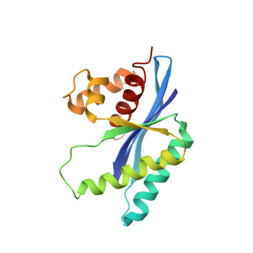
Atomic structure of the RuvC resolvase: a holliday junction-specific endonuclease from E. coli.

Ariyoshi, M.Vassylyev, D.G.Iwasaki, H.Nakamura, H.Shinagawa, H.Morikawa, K.

(1994) Cell 78: 1063-1072

  • DOI: https://doi.org/10.1016/0092-8674(94)90280-1
  • Primary Citation of Related Structures:  
    1HJR

  • PubMed Abstract: 

    The crystal structure of the RuvC protein, a Holliday junction resolvase from E. coli, has been determined at 2.5 A resolution. The enzyme forms a dimer of 19 kDa subunits related by a dyad axis. Together with results from extensive mutational analyses, the refined structure reveals that the catalytic center, comprising four acidic residues, lies at the bottom of a cleft that nicely fits a DNA duplex. The structural features of the dimer, with a 30 A spacing between the two catalytic centers, provide a substantially defined image of the Holliday junction architecture. The folding topology in the vicinity of the catalytic site exhibits a striking similarity to that of RNAase H1 from E. coli.


  • Organizational Affiliation

    Protein Engineering Research Institute, Osaka, Japan.


Macromolecules
Find similar proteins by:  (by identity cutoff)  |  3D Structure
Entity ID: 1
MoleculeChains Sequence LengthOrganismDetailsImage
HOLLIDAY JUNCTION RESOLVASE (RUVC)
A, B, C, D
158Escherichia coliMutation(s): 0 
EC: 3.1.22.4
UniProt
Find proteins for P0A814 (Escherichia coli (strain K12))
Explore P0A814 
Go to UniProtKB:  P0A814
Entity Groups  
Sequence Clusters30% Identity50% Identity70% Identity90% Identity95% Identity100% Identity
UniProt GroupP0A814
Sequence Annotations
Expand
  • Reference Sequence
Experimental Data & Validation

Experimental Data

  • Method: X-RAY DIFFRACTION
  • Resolution: 2.50 Å
  • R-Value Work: 0.157 
  • R-Value Observed: 0.157 
  • Space Group: P 1 21 1
Unit Cell:
Length ( Å )Angle ( ˚ )
a = 72.8α = 90
b = 139.6β = 93
c = 32.4γ = 90
Software Package:
Software NamePurpose
X-PLORmodel building
PROLSQrefinement
X-PLORrefinement
X-PLORphasing

Structure Validation

View Full Validation Report


Entry History 

Deposition Data

Revision History  (Full details and data files)

  • Version 1.0: 1995-02-07
    Type: Initial release
  • Version 1.1: 2008-03-24
    Changes: Version format compliance
  • Version 1.2: 2011-07-13
    Changes: Version format compliance
  • Version 1.3: 2019-07-17
    Changes: Data collection, Other, Refinement description
  • Version 1.4: 2019-08-14
    Changes: Data collection, Refinement description
  • Version 1.5: 2024-02-07
    Changes: Data collection, Database references