Insight Into the Stereochemistry in the Inhibition of Carboxypeptidase a with N-(Hydroxyaminocarbonyl)Phenylalanine: Binding Modes of an Enantiomeric Pair of the Inhibitor to Carboxypeptidase A
Cho, J.H., Kim, D.H., Chung, S.J., Ha, N.-C., Oh, B.-H., Choi, K.Y.(2002) Bioorg Med Chem 10: 2015
- PubMed: 11937361
- DOI: https://doi.org/10.1016/s0968-0896(01)00429-1
- Primary Citation of Related Structures:
1HDQ, 1HDU, 1HEE - PubMed Abstract:
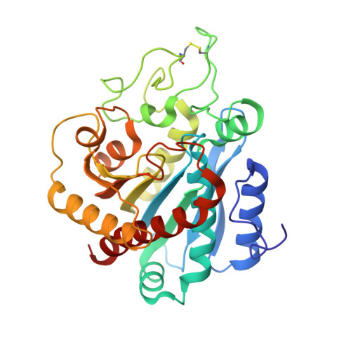
Both D- and L-isomers of N-(hydroxyaminocarbonyl)phenylalanine () were shown to have strong binding affinity towards carboxypeptidase A (CPA) with D- being more potent than its enantiomer by 3-fold (Chung, S. J.; Kim, D. H. Bioorg. Med. Chem. 2001, 9, 185.). In order to understand the reversed stereochemical preference shown in the CPA inhibition, we have solved the crystal structures of CPA complexed with each enantiometer of up to 1.75 A resolution. Inhibitor L- whose stereochemistry belongs to the stereochemical series of substrate binds CPA like substrate does with its carbonyl oxygen coordinating to the active site zinc ion. Its hydroxyl is engaged in hydrogen bonding with the carboxylate of Glu-270. On the other hand, in binding of D- to CPA, its terminal hydroxyl group is involved in interactions with the active site zinc ion and the carboxylate of Glu-270. In both CPA small middle dot complexes, the phenyl ring in is fitted in the substrate recognition pocket at the S(1)' subsite, and the carboxylate of the inhibitors forms bifurcated hydrogen bonds with the guanidinium moiety of Arg-145 and a hydrogen bond with the guanidinium of Arg-127. In the complex of CPA small middle dotD-, the carboxylate of the inhibitor is engaged in hydrogen bonding with the phenolic hydroxyl of the down-positioned Tyr-248. While the L- binding induces a concerted movement of the backbone amino acid residues at the active site, only the downward movement of Tyr-248 was noted when D- binds to CPA.
Organizational Affiliation:
National Research Laboratory for Protein Engineering, Pohang University of Science and Technology, San 31Hyoja-dong, 790-784, Pohang, Republic of Korea.