1HBV

A CHECK ON RATIONAL DRUG DESIGN. CRYSTAL STRUCTURE OF A COMPLEX OF HIV-1 PROTEASE WITH A NOVEL GAMMA-TURN MIMETIC


Experimental Data Snapshot

  • Method: X-RAY DIFFRACTION
  • Resolution: 2.30 Å
  • R-Value Observed: 0.177 

wwPDB Validation   3D Report Full Report


Ligand Structure Quality Assessment 


This is version 1.4 of the entry. See complete history


Literature

A check on rational drug design: crystal structure of a complex of human immunodeficiency virus type 1 protease with a novel gamma-turn mimetic inhibitor.

Hoog, S.S.Zhao, B.Winborne, E.Fisher, S.Green, D.W.DesJarlais, R.L.Newlander, K.A.Callahan, J.F.Moore, M.L.Huffman, W.F.Abdel-Meguid, S.S.

(1995) J Med Chem 38: 3246-3252

  • DOI: https://doi.org/10.1021/jm00017a008
  • Primary Citation of Related Structures:  
    1HBV

  • PubMed Abstract: 

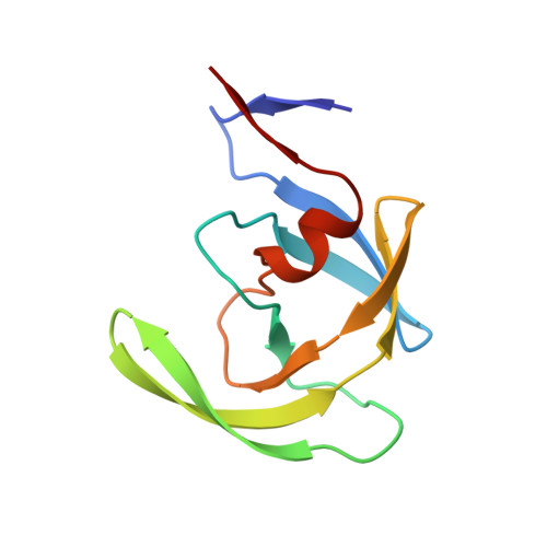
    We have previously reported (Newlander et al., J. Med. Chem. 1993, 36, 2321-2331) the design of human immunodeficiency virus type 1 (HIV-1) protease inhibitors incorporating C7 mimetics that lock three amino acid residues of a peptide sequence into a gamma-turn. The design of one such compound, SB203238, was based on X-ray structures of reduced amide aspartyl protease inhibitors. It incorporates a gamma-turn mimetic in the P2-P1' position, where the carbonyl of the C7 ring is replaced with an sp3 methylene group yielding a constrained reduced amide. It shows competitive inhibition with Ki = 430 nM at pH 6.0. The three-dimensional structure of SB203238 bound to the active site of HIV-1 protease has been determined at 2.3 A resolution by X-ray diffraction and refined to a crystallographic R-factor (R = sigma magnitude of Fo magnitude of - magnitude of Fc magnitude of /sigma magnitude of Fo magnitude of, where Fo and Fc are the observed and calculated structure factor amplitudes, respectively) of 0.177. The inhibitor lies in an extended conformation in the active site; however, because of the constrained geometry of the C7 ring, it maintains fewer hydrogen bonds with the protein than in most other HIV-1 protease-inhibitor complexes. More importantly, the inhibitor binds to the enzyme differently than predicted in its design, by binding with the P2-P1' alpha-carbon atoms shifted by approximately one-half a residue toward the N-terminus from their presumed positions. This study illustrates the importance of structural information in an approach to rational drug design.


  • Organizational Affiliation

    Department of Macromolecular Sciences, SmithKline Beecham Pharmaceuticals, King of Prussia, Pennsylvania 19406, USA.


Macromolecules
Find similar proteins by:  (by identity cutoff)  |  3D Structure
Entity ID: 1
MoleculeChains Sequence LengthOrganismDetailsImage
HIV-1 PROTEASE
A, B
99Human immunodeficiency virus 1Mutation(s): 0 
Gene Names: HIV-1 PROTEASE
UniProt
Find proteins for P03366 (Human immunodeficiency virus type 1 group M subtype B (isolate BH10))
Explore P03366 
Go to UniProtKB:  P03366
Entity Groups  
Sequence Clusters30% Identity50% Identity70% Identity90% Identity95% Identity100% Identity
UniProt GroupP03366
Sequence Annotations
Expand
  • Reference Sequence
Small Molecules
Ligands 1 Unique
IDChains Name / Formula / InChI Key2D Diagram3D Interactions
GAN
Query on GAN

Download Ideal Coordinates CCD File 
C [auth A]2-[3-BENZYL-5-(1-ALANYL-AMINOETHYL)-2,3,6,7-TETRAHYDRO-1H-AZEPIN-1-YL]-1-OXOPROPYL-VALINYL-VALINE-METHYLESTER
C32 H51 N5 O5
AAROLUGLGDXGEZ-CFILSVQKSA-N
Binding Affinity Annotations 
IDSourceBinding Affinity
GAN Binding MOAD:  1HBV Ki: 430 (nM) from 1 assay(s)
PDBBind:  1HBV Ki: 430 (nM) from 1 assay(s)
BindingDB:  1HBV Ki: 437 (nM) from 1 assay(s)
Kd: 427 (nM) from 1 assay(s)
Experimental Data & Validation

Experimental Data

  • Method: X-RAY DIFFRACTION
  • Resolution: 2.30 Å
  • R-Value Observed: 0.177 
  • Space Group: P 61
Unit Cell:
Length ( Å )Angle ( ˚ )
a = 63.4α = 90
b = 63.4β = 90
c = 83.9γ = 120
Software Package:
Software NamePurpose
X-PLORmodel building
PROLSQrefinement
X-PLORrefinement
XENGENdata reduction
X-PLORphasing

Structure Validation

View Full Validation Report


Ligand Structure Quality Assessment 


Entry History 

Deposition Data

Revision History  (Full details and data files)

  • Version 1.0: 1995-07-10
    Type: Initial release
  • Version 1.1: 2008-03-24
    Changes: Version format compliance
  • Version 1.2: 2011-07-13
    Changes: Version format compliance
  • Version 1.3: 2012-02-22
    Changes: Database references
  • Version 1.4: 2024-02-07
    Changes: Data collection, Database references, Derived calculations, Other