Crystal structure of liganded and unliganded forms of bovine plasma retinol-binding protein.
Zanotti, G., Berni, R., Monaco, H.L.(1993) J Biol Chem 268: 10728-10738
- PubMed: 8496140
- Primary Citation of Related Structures:
1HBP, 1HBQ - PubMed Abstract:
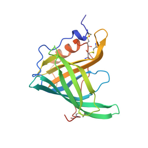
The three-dimensional structures of bovine plasma retinol-binding protein (bRBP) complexed with retinol (space group P2(1)2(1)2(1), a = 46.08, b = 49.12, c = 76.10 A) and of the unliganded protein prepared in vitro by extracting retinol with ethyl ether (space group P2(1)2(1)2(1), a = 46.55, b = 48.97, c = 76.87 A) have been solved at 1.9 and 1.7 A resolution, respectively. The final crystallographic R factors are 0.190 for holobRBP and 0.196 for the unliganded bRBP. The model for the bovine holoprotein is quite similar to that of the human protein, with which it exhibits 92% sequence similarity. The root mean square deviation between the alpha-carbons in the two proteins is 0.31 A. The retinol binding site is almost completely preserved. The loops that surround the opening of the beta-barrel are also particularly conserved, in contrast with the presence of several substitutions in parts of the RBP molecule opposite the opening of the calyx that binds retinol. Despite the fact that unliganded bovine RBP was prepared and crystallized using procedures completely different from those used to obtain the unliganded human RBP, the conformational differences between unliganded and liganded forms of bRBP are almost identical to those found previously between the same forms of human RBP. They mainly involve a few residues in the region extending from amino acid residues 32 to 37. Therefore, similar differences are very likely to exist between holoRBP and the physiologically occurring apoprotein. A not yet identified electron density, different in shape and orientation from retinol, also occupies the central cavity of the beta-barrel in the unliganded bRBP, as found for unliganded human RBP. The functional consequences of the conformational change induced by the removal of retinol on the interaction between RBP and transthyretin, coupled with the conservation of the entrance loops of the beta-barrel in mammalian RBPs, are consistent with their participation in molecular interactions.
Organizational Affiliation:
Department of Organic Chemistry, University of Padova, Italy.