1H96

recombinant mouse L-chain ferritin


Experimental Data Snapshot

  • Method: X-RAY DIFFRACTION
  • Resolution: 1.60 Å
  • R-Value Free: 0.220 
  • R-Value Work: 0.160 

wwPDB Validation   3D Report Full Report


This is version 1.4 of the entry. See complete history


Literature

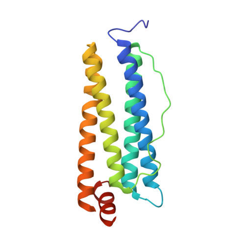
Structure of Mouse L-Chain Ferritin at 1.6 A Resolution

Granier, T.Gallois, B.D'Estaintot, B.L.Dautant, A.Chevalier, J.M.Mellado, J.M.Beaumont, C.Santambrogio, P.Arosio, P.Precigoux, G.

(2001) Acta Crystallogr D Biol Crystallogr 57: 1491

  • DOI: https://doi.org/10.1107/s0907444901008897
  • Primary Citation of Related Structures:  
    1H96

  • PubMed Abstract: 

    Cubic F432 crystals of recombinant mouse L-chain apoferritin were obtained by the hanging-drop technique with ammonium sulfate and cadmium sulfate as precipitants. The structure was refined to 2.1 and 1.6 A resolution from data obtained at room temperature and under cryogenic conditions, respectively. The structure of an eight-amino-acid loop insertion in the mouse sequence is found to be highly disordered both at room temperature and at low temperature.


  • Organizational Affiliation

    Unité de Biophysique Structurale, UMR CNRS 5471, Université Bordeaux I, Bâtiment B8, Avenue des Facultés, 33405 Talence CEDEX, France. t.granier@ubs.u-bordeaux.fr


Macromolecules
Find similar proteins by:  (by identity cutoff)  |  3D Structure
Entity ID: 1
MoleculeChains Sequence LengthOrganismDetailsImage
FERRITIN LIGHT CHAIN 1182Mus musculusMutation(s): 0 
UniProt & NIH Common Fund Data Resources
Find proteins for P29391 (Mus musculus)
Explore P29391 
Go to UniProtKB:  P29391
IMPC:  MGI:95589
Entity Groups  
Sequence Clusters30% Identity50% Identity70% Identity90% Identity95% Identity100% Identity
UniProt GroupP29391
Sequence Annotations
Expand
  • Reference Sequence
Small Molecules
Ligands 2 Unique
IDChains Name / Formula / InChI Key2D Diagram3D Interactions
CD
Query on CD

Download Ideal Coordinates CCD File 
B [auth A]
C [auth A]
D [auth A]
E [auth A]
F [auth A]
B [auth A],
C [auth A],
D [auth A],
E [auth A],
F [auth A],
G [auth A],
H [auth A],
I [auth A],
J [auth A]
CADMIUM ION
Cd
WLZRMCYVCSSEQC-UHFFFAOYSA-N
SO4
Query on SO4

Download Ideal Coordinates CCD File 
K [auth A]SULFATE ION
O4 S
QAOWNCQODCNURD-UHFFFAOYSA-L
Experimental Data & Validation

Experimental Data

  • Method: X-RAY DIFFRACTION
  • Resolution: 1.60 Å
  • R-Value Free: 0.220 
  • R-Value Work: 0.160 
  • Space Group: F 4 3 2
Unit Cell:
Length ( Å )Angle ( ˚ )
a = 180.82α = 90
b = 180.82β = 90
c = 180.82γ = 90
Software Package:
Software NamePurpose
REFMACrefinement
MOSFLMdata reduction
SCALAdata scaling
AMoREphasing

Structure Validation

View Full Validation Report


Entry History 

Revision History  (Full details and data files)

  • Version 1.0: 2001-03-06
    Type: Initial release
  • Version 1.1: 2011-05-07
    Changes: Version format compliance
  • Version 1.2: 2011-07-13
    Changes: Version format compliance
  • Version 1.3: 2019-05-08
    Changes: Data collection, Experimental preparation, Other
  • Version 1.4: 2023-12-13
    Changes: Data collection, Database references, Derived calculations, Other, Refinement description