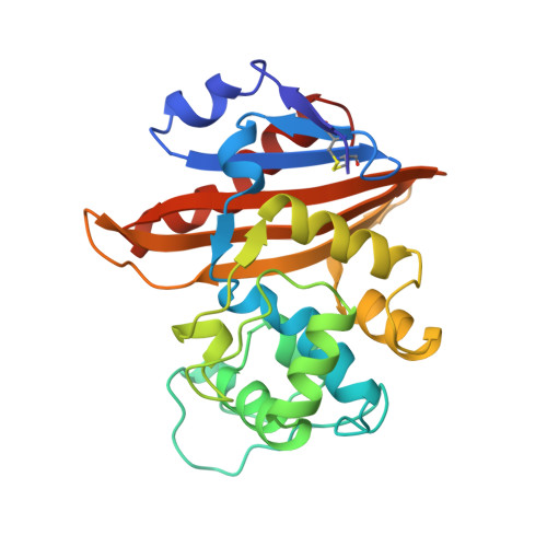
Crystal Structures of the Class D B-Lactamase Oxa-13 in the Native Form and in Complex with Meropenem
Pernot, L., Frenois, F., Rybkine, T., L'Hermite, G., Petrella, S., Delettre, J., Jarlier, V., Collatz, E., Sougakoff, W.(2001) J Mol Biol 310: 859
- PubMed: 11453693
- DOI: https://doi.org/10.1006/jmbi.2001.4805
- Primary Citation of Related Structures:
1H8Y, 1H8Z - PubMed Abstract:
The therapeutic problems posed by class D beta-lactamases, a family of serine enzymes that hydrolyse beta-lactam antibiotics following an acylation-deacylation mechanism, are increased by the very low level of sensitivity of these enzymes to beta-lactamase inhibitors. To gain structural and mechanistic insights to aid the design of new inhibitors, we have determined the crystal structure of OXA-13 from Pseudomonas aeruginosa in the apo form and in complex with the carbapenem meropenem. The native form consisted of a dimer displaying an overall organisation similar to that found in the closely related enzyme OXA-10. In the acyl-enzyme complex, the positioning of the antibiotic appeared to be ensured mainly by (i) the covalent acyl bond and (ii) a strong salt-bridge involving the carboxylate moiety of the drug. Comparison of the structures of OXA-13 in the apo form and in complex with meropenem revealed an unsuspected flexibility in the region of the essential serine 115 residue, with possible consequences for the catalytic properties of the enzyme. In the apo form, the Ser115 side-chain is oriented outside the active site, whereas the general base Lys70 adopts a conformation that seems to be incompatible with the activation of the catalytic water molecule required for the deacylation step. In the OXA-13:meropenem complex, a 3.5 A movement of the backbone of the 114-116 loop towards the side-chain of Lys70 was observed, which seems to be driven by a displacement of the neighbouring 91-104 loop and which results in the repositioning of the side-chain hydroxyl group of Ser115 toward the catalytic centre. Concomitantly, the side-chain of Lys70 is forced to curve in the direction of the deacylating water molecule, which is then strongly bound and activated by this residue. However, a distance of ca 5 A separates the catalytic water molecule from the acyl carbonyl group of meropenem, a structural feature that accounts for the inhibition of OXA-13 by this drug. Finally, the low level of penicillinase activity revealed by the kinetic analysis of OXA-13 could be related to the specific presence in position 73 of a serine residue located close to the general base Lys70, which results in a decrease of the number of hydrogen-bonding interactions stabilising the catalytic water molecule.
Organizational Affiliation:
Laboratoire de Recherche Moléculaire sur les Antibiotiques (LRMA), Facultés de Médecine Pitié-Salpêtrière and Broussais-Hôtel Dieu, Université Pierre et Marie Curie, 91 bd de l'Hôpital, Paris cedex 13, 75634, France.