Structural Basis for the Thioredoxin-Like Activity Profile of the Glutaredoxin-Like Nrdh-Redoxin from Escherichia Coli
Stehr, M., Schneider, G., Aslund, F., Holmgren, A., Lindqvist, Y.(2001) J Biol Chem 276: 35836
- PubMed: 11441020
- DOI: https://doi.org/10.1074/jbc.M105094200
- Primary Citation of Related Structures:
1H75 - PubMed Abstract:
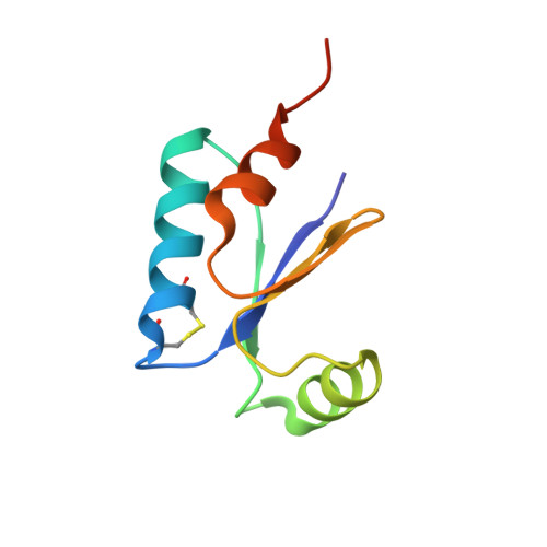
NrdH-redoxin is a representative of a class of small redox proteins that contain a conserved CXXC motif and are characterized by a glutaredoxin-like amino acid sequence and thioredoxin-like activity profile. The crystal structure of recombinant Escherichia coli NrdH-redoxin in the oxidized state has been determined at 1.7 A resolution by multiwavelength anomalous diffraction. NrdH-redoxin belongs to the thioredoxin superfamily and is structurally most similar to E. coli glutaredoxin 3 and phage T4 glutaredoxin. The angle between the C-terminal helix alpha3 and strand beta4, which differs between thioredoxin and glutaredoxin, has an intermediate value in NrdH-redoxin. The orientation of this helix is to a large extent determined by an extended hydrogen-bond network involving the highly conserved sequence motif (61)WSGFRP(D/E)(67), which is unique to this subclass of the thioredoxin superfamily. Residues that bind glutathione in glutaredoxins are in general not conserved in NrdH-redoxin, and no glutathione-binding cleft is present. Instead, NrdH-redoxin contains a wide hydrophobic pocket at the surface, similar to thioredoxin. Modeling studies suggest that NrdH-redoxin can interact with E. coli thioredoxin reductase at this pocket and also via a loop that is complementary to a crevice in the reductase in a similar manner as observed in the E. coli thioredoxin-thioredoxin reductase complex.
Organizational Affiliation:
Division of Molecular Structural Biology and the Medical Nobel Institute for Biochemistry, Department of Medical Biochemistry and Biophysics, Karolinska Institute, Stockholm, S-171 77 Sweden.