1H70

DDAH FROM PSEUDOMONAS AERUGINOSA. C249S MUTANT COMPLEXED WITH CITRULLINE


Experimental Data Snapshot

  • Method: X-RAY DIFFRACTION
  • Resolution: 1.80 Å
  • R-Value Free: 0.218 
  • R-Value Work: 0.190 
  • R-Value Observed: 0.191 

wwPDB Validation   3D Report Full Report


This is version 2.0 of the entry. See complete history


Literature

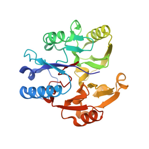
Structural insights into the hydrolysis of cellular nitric oxide synthase inhibitors by dimethylarginine dimethylaminohydrolase.

Murray-Rust, J.Leiper, J.McAlister, M.Phelan, J.Tilley, S.Santa Maria, J.Vallance, P.McDonald, N.

(2001) Nat Struct Biol 8: 679-683

  • DOI: https://doi.org/10.1038/90387
  • Primary Citation of Related Structures:  
    1H70

  • PubMed Abstract: 

    Nitric oxide synthase is inhibited by asymmetric NG-methylated derivatives of arginine whose cellular levels are controlled in part by dimethylarginine dimethylaminohydrolase (DDAH, EC 3.5.3.18). Levels of asymmetric NG,NG-dimethylarginine (ADMA) are known to correlate with certain disease states. Here, the first structure of a DDAH shows an unexpected similarity to arginine:glycine amidinotransferase (EC 2.1.4.1) and arginine deiminase (EC 3.5.3.6), thus defining a superfamily of arginine-modifying enzymes. The identification of a Cys-His-Glu catalytic triad and the structures of a Cys to Ser point mutant bound to both substrate and product suggest a reaction mechanism. Comparison of the ADMA-DDAH and arginine-amidinotransferase complexes reveals a dramatic rotation of the substrate that effectively maintains the orientation of the scissile bond of the substrate with respect to the catalytic residues. The DDAH structure will form a basis for the rational design of selective inhibitors, which are of potential use in modulating NO synthase activity in pathological settings.


  • Organizational Affiliation

    School of Crystallography, Birkbeck, Malet Street, London WC1E 7HX, UK.


Macromolecules
Find similar proteins by:  (by identity cutoff)  |  3D Structure
Entity ID: 1
MoleculeChains Sequence LengthOrganismDetailsImage
NG, NG-DIMETHYLARGININE DIMETHYLAMINOHYDROLASE255Pseudomonas aeruginosaMutation(s): 1 
EC: 3.5.3.18
UniProt
Find proteins for Q9I4E3 (Pseudomonas aeruginosa (strain ATCC 15692 / DSM 22644 / CIP 104116 / JCM 14847 / LMG 12228 / 1C / PRS 101 / PAO1))
Explore Q9I4E3 
Go to UniProtKB:  Q9I4E3
Entity Groups  
Sequence Clusters30% Identity50% Identity70% Identity90% Identity95% Identity100% Identity
UniProt GroupQ9I4E3
Sequence Annotations
Expand
  • Reference Sequence
Small Molecules
Ligands 1 Unique
IDChains Name / Formula / InChI Key2D Diagram3D Interactions
CIR
Query on CIR

Download Ideal Coordinates CCD File 
B [auth A]CITRULLINE
C6 H13 N3 O3
RHGKLRLOHDJJDR-BYPYZUCNSA-N
Experimental Data & Validation

Experimental Data

  • Method: X-RAY DIFFRACTION
  • Resolution: 1.80 Å
  • R-Value Free: 0.218 
  • R-Value Work: 0.190 
  • R-Value Observed: 0.191 
  • Space Group: C 1 2 1
Unit Cell:
Length ( Å )Angle ( ˚ )
a = 69.814α = 90
b = 61.798β = 95.15
c = 54.618γ = 90
Software Package:
Software NamePurpose
REFMACrefinement
MOSFLMdata reduction
SCALAdata scaling
AMoREphasing

Structure Validation

View Full Validation Report


Entry History 

Deposition Data

Revision History  (Full details and data files)

  • Version 1.0: 2001-08-02
    Type: Initial release
  • Version 1.1: 2011-05-08
    Changes: Version format compliance
  • Version 1.2: 2011-07-13
    Changes: Version format compliance
  • Version 1.3: 2018-02-28
    Changes: Advisory, Database references
  • Version 1.4: 2019-05-08
    Changes: Data collection, Experimental preparation
  • Version 2.0: 2023-11-15
    Changes: Advisory, Atomic model, Data collection, Database references, Derived calculations