1H6E

MU2 ADAPTIN SUBUNIT (AP50) OF AP2 ADAPTOR (SECOND DOMAIN), COMPLEXED WITH CTLA-4 INTERNALIZATION PEPTIDE TTGVYVKMPPT


Experimental Data Snapshot

  • Method: X-RAY DIFFRACTION
  • Resolution: 3.60 Å
  • R-Value Free: 0.317 
  • R-Value Work: 0.278 

wwPDB Validation   3D Report Full Report


This is version 2.1 of the entry. See complete history


Literature

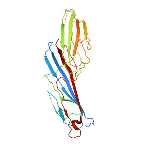
Study of the Interaction of the Medium Chain Mu2 Subunit of the Clathrin-Associated Adapter Protein Complex 2 with Cytotoxic T-Lymphocyte Antigen 4 and Cd28

Follows, E.R.Mcpheat, J.C.Minshull, C.Moore, N.C.Pauptit, R.A.Rowsell, S.Stacey, C.L.Stanway, J.J.Taylor, I.W.Abbott, W.M.

(2001) Biochem J 359: 427

  • DOI: https://doi.org/10.1042/0264-6021:3590427
  • Primary Citation of Related Structures:  
    1H6E

  • PubMed Abstract: 

    The medium chain mu 2 subunit (AP50) of the clathrin-associated adapter protein complex 2 (AP-2) interacts specifically with the tyrosine-based signals of several integral membrane proteins through the consensus sequence YXXPhi, where X can be any residue and Phi is a large hydrophobic residue. Using surface plasmon resonance combined with structural information, we have analysed the interaction of AP50 with peptides derived from the cytoplasmic tail of cytotoxic T-lymphocyte antigen 4 (CTLA-4). The crystal structure of AP50 in complex with a CTLA-4-derived peptide was determined to 3.6 A (1 A=0.1 nm) resolution. The binding domain of AP50 (residues 164-435) was expressed in Escherichia coli and purified. In agreement with previous reports, the AP50 domain bound to residues 152-174 of CTLA-4, but not to the same peptide that was phosphorylated at the single tyrosine residue (position 165). The interaction exhibited fast kinetics with rapid on and off rates and a K(d) of 0.7 microM. In order to further understand why AP50 binds to CTLA-4, but not to the homologous receptor CD28, a comparison of binding of AP50 with five peptides with single changes in and around the YXXPhi motif to the equivalent residues of CD28 was made. T162H greatly reduced binding, whereas T161L had little effect. Mutations G163S, V164D and K167N all exhibited reduced binding. Modelling of the single amino acid changes using structural information, was in broad agreement with the binding data, demonstrating that residues outside of the YXXPhi motif are also important in the interaction of membrane proteins with AP50.


  • Organizational Affiliation

    Enabling Science and Technology, Biology Department, AstraZeneca, Mereside, Alderley Park, Macclesfield, Cheshire SK10 4TG, UK.


Macromolecules
Find similar proteins by:  (by identity cutoff)  |  3D Structure
Entity ID: 1
MoleculeChains Sequence LengthOrganismDetailsImage
CLATHRIN COAT ASSEMBLY PROTEIN AP50288Homo sapiensMutation(s): 0 
UniProt & NIH Common Fund Data Resources
Find proteins for Q96CW1 (Homo sapiens)
Explore Q96CW1 
Go to UniProtKB:  Q96CW1
PHAROS:  Q96CW1
GTEx:  ENSG00000161203 
Entity Groups  
Sequence Clusters30% Identity50% Identity70% Identity90% Identity95% Identity100% Identity
UniProt GroupQ96CW1
Sequence Annotations
Expand
  • Reference Sequence

Find similar proteins by:  Sequence   |   3D Structure  

Entity ID: 2
MoleculeChains Sequence LengthOrganismDetailsImage
CYTOTOXIC T-LYMPHOCYTE PROTEIN 4B [auth P]11Homo sapiensMutation(s): 0 
UniProt & NIH Common Fund Data Resources
Find proteins for P16410 (Homo sapiens)
Explore P16410 
Go to UniProtKB:  P16410
PHAROS:  P16410
GTEx:  ENSG00000163599 
Entity Groups  
Sequence Clusters30% Identity50% Identity70% Identity90% Identity95% Identity100% Identity
UniProt GroupP16410
Sequence Annotations
Expand
  • Reference Sequence
Experimental Data & Validation

Experimental Data

  • Method: X-RAY DIFFRACTION
  • Resolution: 3.60 Å
  • R-Value Free: 0.317 
  • R-Value Work: 0.278 
  • Space Group: P 64
Unit Cell:
Length ( Å )Angle ( ˚ )
a = 126.779α = 90
b = 126.779β = 90
c = 74.161γ = 120
Software Package:
Software NamePurpose
CNXrefinement
MOSFLMdata reduction
SCALAdata scaling
CCP4phasing

Structure Validation

View Full Validation Report


Entry History 

Deposition Data

Revision History  (Full details and data files)

  • Version 1.0: 2001-11-28
    Type: Initial release
  • Version 1.1: 2016-12-21
    Changes: Data collection, Derived calculations, Refinement description, Source and taxonomy, Version format compliance
  • Version 2.0: 2019-05-29
    Changes: Atomic model, Data collection, Experimental preparation, Other
  • Version 2.1: 2023-12-13
    Changes: Data collection, Database references, Other, Refinement description