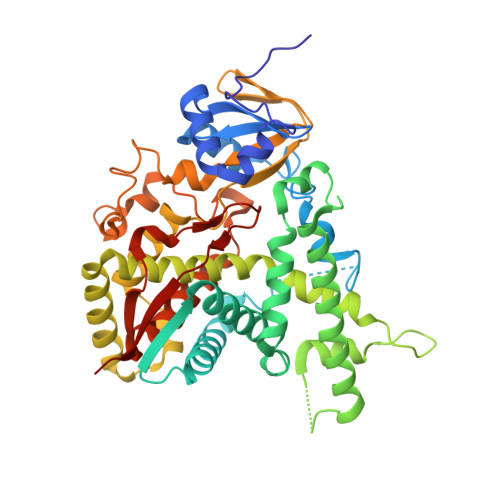
1H5Z

CYTOCHROME P450 14 ALPHA-STEROL DEMETHYLASE (CYP51) FROM MYCOBACTERIUM TUBERCULOSIS IN FERRIC LOW-SPIN STATE


Experimental Data Snapshot

  • Method: X-RAY DIFFRACTION
  • Resolution: 2.05 Å
  • R-Value Free: 0.236 
  • R-Value Work: 0.185 
  • R-Value Observed: 0.185 

wwPDB Validation   3D Report Full Report


Ligand Structure Quality Assessment 


This is version 1.4 of the entry. See complete history


Literature

Estriol Bound and Ligand-Free Structures of Sterol 14Alpha-Demethylase.

Podust, L.M.Yermalitskaya, L.V.Lepesheva, G.I.Podust, V.N.Dalmasso, E.A.Waterman, M.R.

(2004) Structure 12: 1937

  • DOI: https://doi.org/10.1016/j.str.2004.08.009
  • Primary Citation of Related Structures:  
    1H5Z, 1X8V

  • PubMed Abstract: 

    Sterol 14alpha-demethylases (CYP51) are essential enzymes in sterol biosynthesis in eukaryotes and drug targets in antifungal therapy. Here, we report CYP51 structures in ligand-free and estriol bound forms. Using estriol as a probe, we determined orientation of the substrate in the active site, elucidated protein contacts with the invariant 3beta-hydroxy group of a sterol, and identified F78 as a key discriminator between 4alpha-methylated and 4alpha,beta-dimethylated substrates. Analysis of CYP51 dynamics revealed that the C helix undergoes helix-coil transition upon binding and dissociation of a ligand. Loss of helical structure of the C helix in the ligand-free form results in an unprecedented opening of the substrate binding site. Upon binding of estriol, the BC loop loses contacts with molecular surface and tends to adopt a closed conformation. A mechanism for azole resistance in the yeast pathogen Candida albicans associated with mutations in the ERG11 gene encoding CYP51 is suggested based on CYP51 protein dynamics.


  • Organizational Affiliation

    Department of Biochemistry, Vanderbilt University School of Medicine, Nashville, TN 37232, USA. larissa.m.podust@vanderbilt.edu


Macromolecules
Find similar proteins by:  (by identity cutoff)  |  3D Structure
Entity ID: 1
MoleculeChains Sequence LengthOrganismDetailsImage
CYTOCHROME P450 51455Mycobacterium tuberculosisMutation(s): 0 
Gene Names: CYP51RV0764CMTCY369.09C
EC: 1.14.13.70
UniProt
Find proteins for P9WPP9 (Mycobacterium tuberculosis (strain ATCC 25618 / H37Rv))
Explore P9WPP9 
Go to UniProtKB:  P9WPP9
Entity Groups  
Sequence Clusters30% Identity50% Identity70% Identity90% Identity95% Identity100% Identity
UniProt GroupP9WPP9
Sequence Annotations
Expand
  • Reference Sequence
Small Molecules
Ligands 2 Unique
IDChains Name / Formula / InChI Key2D Diagram3D Interactions
HEM
Query on HEM

Download Ideal Coordinates CCD File 
B [auth A]PROTOPORPHYRIN IX CONTAINING FE
C34 H32 Fe N4 O4
KABFMIBPWCXCRK-RGGAHWMASA-L
FE2
Query on FE2

Download Ideal Coordinates CCD File 
C [auth A]FE (II) ION
Fe
CWYNVVGOOAEACU-UHFFFAOYSA-N
Experimental Data & Validation

Experimental Data

  • Method: X-RAY DIFFRACTION
  • Resolution: 2.05 Å
  • R-Value Free: 0.236 
  • R-Value Work: 0.185 
  • R-Value Observed: 0.185 
  • Space Group: P 21 21 21
Unit Cell:
Length ( Å )Angle ( ˚ )
a = 46.489α = 90
b = 84.827β = 90
c = 110.025γ = 90
Software Package:
Software NamePurpose
CNSrefinement
DENZOdata reduction
SCALEPACKdata scaling
CNSphasing

Structure Validation

View Full Validation Report


Ligand Structure Quality Assessment 


Entry History 

Deposition Data

Revision History  (Full details and data files)

  • Version 1.0: 2003-10-03
    Type: Initial release
  • Version 1.1: 2015-04-15
    Changes: Database references, Derived calculations, Non-polymer description, Other, Version format compliance
  • Version 1.2: 2015-08-05
    Changes: Derived calculations, Other, Source and taxonomy
  • Version 1.3: 2019-05-08
    Changes: Data collection, Experimental preparation, Other
  • Version 1.4: 2023-12-13
    Changes: Data collection, Database references, Derived calculations, Other, Refinement description