1H5V

Thiopentasaccharide complex of the endoglucanase Cel5A from Bacillus agaradharens at 1.1 A resolution in the tetragonal crystal form


Experimental Data Snapshot

  • Method: X-RAY DIFFRACTION
  • Resolution: 1.10 Å
  • R-Value Free: 0.138 
  • R-Value Work: 0.120 
  • R-Value Observed: 0.121 

wwPDB Validation   3D Report Full Report


This is version 2.1 of the entry. See complete history


Literature

Atomic Resolution Structure of Endoglucanase Cel5A in Complex with Methyl 4,4II,4III,4Iv-Tetrathio-Alpha-Cellopentoside Highlights the Alternative Binding Modes Targeted by Substrate Mimics

Varrot, A.Schulein, M.Fruchard, S.Driguez, H.Davies, G.J.

(2001) Acta Crystallogr D Biol Crystallogr 57: 1739

  • DOI: https://doi.org/10.1107/s0907444901013993
  • Primary Citation of Related Structures:  
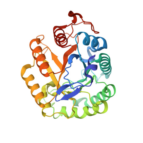
    1H5V

  • PubMed Abstract: 

    Many three-dimensional structures of retaining beta-D-glycoside hydrolases have been determined, yet oligosaccharide complexes in which the ligand spans the catalytic centre are rare. Those that have been reported so far have revealed two modes of binding: those in which the substrate adopts a distorted skew-boat or envelope conformation in the -1 subsite, reflecting the distortion observed during the catalytic cycle, and those which bypass the true catalytic centre and thus lie in a non-productive manner across the -1 subsite. The three-dimensional structure of a retaining endocellulase, Bacillus agaradhaerens Cel5A, in complex with methyl 4,4(II),4(III),4(IV)-tetrathio-alpha-cellopentoside falls into this latter category. The 1.1 A structure reveals the binding of five pyranosides, all in the (4)C(1) chair conformation, occupying the -3, -2, +1 and +2 subsites whilst evading the catalytic machinery located in the true -1 subsite. Such binding is in marked contrast to the structure of another retaining endocellulase, the Fusarium oxysporum Cel7B, the identical ligand in which displayed a distorted skew-boat conformation at the active centre. These two binding modes may reflect different steps in the binding and catalytic process.


  • Organizational Affiliation

    Structural Biology Laboratory, Department of Chemistry, University of York, Heslington, York Y010 5DD, England.


Macromolecules
Find similar proteins by:  (by identity cutoff)  |  3D Structure
Entity ID: 1
MoleculeChains Sequence LengthOrganismDetailsImage
ENDOGLUCANASE 5A304Salipaludibacillus agaradhaerensMutation(s): 0 
EC: 3.2.1.4
UniProt
Find proteins for O85465 (Salipaludibacillus agaradhaerens)
Explore O85465 
Go to UniProtKB:  O85465
Entity Groups  
Sequence Clusters30% Identity50% Identity70% Identity90% Identity95% Identity100% Identity
UniProt GroupO85465
Sequence Annotations
Expand
  • Reference Sequence
Oligosaccharides

Help

Entity ID: 2
MoleculeChains Length2D Diagram Glycosylation3D Interactions
alpha-D-glucopyranose-(1-4)-1,4-dithio-beta-D-glucopyranose-(1-4)-4-thio-beta-D-glucopyranose-(1-4)-4-thio-beta-D-glucopyranose-(1-4)-methyl 4-thio-alpha-D-glucopyranoside
B
5N/AN/A
Glycosylation Resources
GlyTouCan:  G68848DV
GlyCosmos:  G68848DV
Small Molecules
Ligands 2 Unique
IDChains Name / Formula / InChI Key2D Diagram3D Interactions
CA
Query on CA

Download Ideal Coordinates CCD File 
C [auth A]
D [auth A]
E [auth A]
F [auth A]
G [auth A]
C [auth A],
D [auth A],
E [auth A],
F [auth A],
G [auth A],
H [auth A],
I [auth A],
J [auth A],
K [auth A],
L [auth A]
CALCIUM ION
Ca
BHPQYMZQTOCNFJ-UHFFFAOYSA-N
NA
Query on NA

Download Ideal Coordinates CCD File 
M [auth A],
N [auth A]
SODIUM ION
Na
FKNQFGJONOIPTF-UHFFFAOYSA-N
Experimental Data & Validation

Experimental Data

  • Method: X-RAY DIFFRACTION
  • Resolution: 1.10 Å
  • R-Value Free: 0.138 
  • R-Value Work: 0.120 
  • R-Value Observed: 0.121 
  • Space Group: P 43 21 2
Unit Cell:
Length ( Å )Angle ( ˚ )
a = 75.169α = 90
b = 75.169β = 90
c = 135.471γ = 90
Software Package:
Software NamePurpose
REFMACrefinement
DENZOdata reduction
SCALEPACKdata scaling

Structure Validation

View Full Validation Report


Entry History 

Deposition Data

Revision History  (Full details and data files)

  • Version 1.0: 2002-05-23
    Type: Initial release
  • Version 1.1: 2011-05-08
    Changes: Version format compliance
  • Version 1.2: 2011-07-13
    Changes: Version format compliance
  • Version 1.3: 2018-10-24
    Changes: Data collection, Source and taxonomy
  • Version 2.0: 2020-07-29
    Type: Remediation
    Reason: Carbohydrate remediation
    Changes: Advisory, Atomic model, Data collection, Derived calculations, Structure summary
  • Version 2.1: 2023-12-13
    Changes: Data collection, Database references, Refinement description, Structure summary