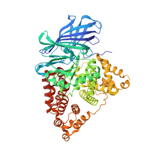
Leukotriene A4 Hydrolase/Aminopeptidase, Glutamate 271 is a Catalyticresidue with Specific Roles in Two Distinct Enzyme Mechanisms
Rudberg, P.C., Tholander, F., Thunnissen, M.M.G.M., Haeggstrom, J.Z.(2002) J Biol Chem 277: 1398
- PubMed: 11675384
- DOI: https://doi.org/10.1074/jbc.M106577200
- Primary Citation of Related Structures:
1H19 - PubMed Abstract:
Leukotriene A(4) hydrolase/aminopeptidase is a bifunctional zinc metalloenzyme that converts the fatty acid epoxide leukotriene A(4) into leukotriene B(4), a potent chemoattractant and immune-modulating lipid mediator. Recently, the structure of leukotriene A(4) hydrolase revealed that Glu-271, which belongs to a conserved GXMEN motif in the M1 family of zinc peptidases, and Gln-136 are located at the active site. Here we report that mutagenetic replacements of Glu-271, but not Gln-136, abrogate both catalytic activities of leukotriene A(4) hydrolase. Furthermore, the 2.1 A crystal structure of [E271Q]leukotriene A(4) hydrolase revealed minimal conformational changes that could not explain the loss of enzyme function. We propose that the carboxylate of Glu-271 participates in an acid-induced opening of the epoxide moiety of leukotriene A(4) and formation of a carbocation intermediate. Moreover, Glu-271 appears to act as an N-terminal recognition site and may potentially stabilize the transition-state during turnover of peptides, a property that most likely pertains to all members of the M1 family of zinc aminopeptidases. Hence, Glu-271 is a unique example of an amino acid, which has dual and separate functions in two different catalytic reactions, involving lipid and peptide substrates, respectively.
Organizational Affiliation:
Department of Medical Biochemistry and Biophysics, Division of Chemistry II, Karolinska Institutet, S-171 77 Stockholm, Sweden.