1GZG

Complex of a Mg2-dependent porphobilinogen synthase from Pseudomonas aeruginosa (mutant D139N) with 5-fluorolevulinic acid


Experimental Data Snapshot

  • Method: X-RAY DIFFRACTION
  • Resolution: 1.66 Å
  • R-Value Free: 0.198 
  • R-Value Work: 0.175 

wwPDB Validation   3D Report Full Report


This is version 2.2 of the entry. See complete history


Literature

Structure of porphobilinogen synthase from Pseudomonas aeruginosa in complex with 5-fluorolevulinic acid suggests a double Schiff base mechanism.

Frere, F.Schubert, W.D.Stauffer, F.Frankenberg, N.Neier, R.Jahn, D.Heinz, D.W.

(2002) J Mol Biol 320: 237-247

  • DOI: https://doi.org/10.1016/S0022-2836(02)00472-2
  • Primary Citation of Related Structures:  
    1GZG

  • PubMed Abstract: 

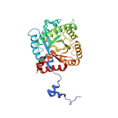
    All natural tetrapyrroles, including hemes, chlorophylls and vitamin B12, share porphobilinogen (PBG) as a common precursor. Porphobilinogen synthase (PBGS) synthesizes PBG through the asymmetric condensation of two molecules of aminolevulinic acid (ALA). Crystal structures of PBGS from various sources confirm the presence of two distinct binding sites for each ALA molecule, termed A and P. We have solved the structure of the active-site variant D139N of the Mg2+-dependent PBGS from Pseudomonas aeruginosa in complex with the inhibitor 5-fluorolevulinic acid at high resolution. Uniquely, full occupancy of both substrate binding sites each by a single substrate-like molecule was observed. Both inhibitor molecules are covalently bound to two conserved, active-site lysine residues, Lys205 and Lys260, through Schiff bases. The active site now also contains a monovalent cation that may critically enhance enzymatic activity. Based on these structural data, we postulate a catalytic mechanism for P. aeruginosa PBGS initiated by a C-C bond formation between A and P-side ALA, followed by the formation of the intersubstrate Schiff base yielding the product PBG.


  • Organizational Affiliation

    Institute of Microbiology, Technical University Braunschweig, Spielmannstrasse 7, D-38106 Braunschweig, Germany.


Macromolecules
Find similar proteins by:  (by identity cutoff)  |  3D Structure
Entity ID: 1
MoleculeChains Sequence LengthOrganismDetailsImage
DELTA-AMINOLEVULINIC ACID DEHYDRATASE
A, B
337Pseudomonas aeruginosaMutation(s): 1 
EC: 4.2.1.24
UniProt
Find proteins for Q59643 (Pseudomonas aeruginosa (strain ATCC 15692 / DSM 22644 / CIP 104116 / JCM 14847 / LMG 12228 / 1C / PRS 101 / PAO1))
Explore Q59643 
Go to UniProtKB:  Q59643
Entity Groups  
Sequence Clusters30% Identity50% Identity70% Identity90% Identity95% Identity100% Identity
UniProt GroupQ59643
Sequence Annotations
Expand
  • Reference Sequence
Small Molecules
Ligands 5 Unique
IDChains Name / Formula / InChI Key2D Diagram3D Interactions
LAF
Query on LAF

Download Ideal Coordinates CCD File 
C [auth A],
D [auth A],
H [auth B],
I [auth B]
5-FLUOROLEVULINIC ACID
C5 H7 F O3
MBVLGMJBSFUHKW-UHFFFAOYSA-N
SO4
Query on SO4

Download Ideal Coordinates CCD File 
G [auth A],
N [auth B]
SULFATE ION
O4 S
QAOWNCQODCNURD-UHFFFAOYSA-L
K
Query on K

Download Ideal Coordinates CCD File 
J [auth B],
K [auth B]
POTASSIUM ION
K
NPYPAHLBTDXSSS-UHFFFAOYSA-N
MG
Query on MG

Download Ideal Coordinates CCD File 
E [auth A],
L [auth B]
MAGNESIUM ION
Mg
JLVVSXFLKOJNIY-UHFFFAOYSA-N
NA
Query on NA

Download Ideal Coordinates CCD File 
F [auth A],
M [auth B]
SODIUM ION
Na
FKNQFGJONOIPTF-UHFFFAOYSA-N
Binding Affinity Annotations 
IDSourceBinding Affinity
LAF PDBBind:  1GZG IC50: 9.00e+5 (nM) from 1 assay(s)
Experimental Data & Validation

Experimental Data

  • Method: X-RAY DIFFRACTION
  • Resolution: 1.66 Å
  • R-Value Free: 0.198 
  • R-Value Work: 0.175 
  • Space Group: P 4 21 2
Unit Cell:
Length ( Å )Angle ( ˚ )
a = 127.131α = 90
b = 127.131β = 90
c = 86.145γ = 90
Software Package:
Software NamePurpose
REFMACrefinement
DENZOdata reduction
SCALEPACKdata scaling
CNSphasing

Structure Validation

View Full Validation Report


Entry History 

Deposition Data

Revision History  (Full details and data files)

  • Version 1.0: 2002-06-27
    Type: Initial release
  • Version 1.1: 2011-05-08
    Changes: Version format compliance
  • Version 1.2: 2011-07-13
    Changes: Version format compliance
  • Version 2.0: 2018-11-21
    Changes: Atomic model, Data collection, Database references, Derived calculations, Structure summary
  • Version 2.1: 2019-05-08
    Changes: Data collection, Derived calculations, Experimental preparation
  • Version 2.2: 2023-12-13
    Changes: Data collection, Database references, Derived calculations, Refinement description