1GWZ

CRYSTAL STRUCTURE OF THE CATALYTIC DOMAIN OF THE PROTEIN TYROSINE PHOSPHATASE SHP-1


Experimental Data Snapshot

  • Method: X-RAY DIFFRACTION
  • Resolution: 2.50 Å
  • R-Value Free: 0.293 
  • R-Value Work: 0.209 
  • R-Value Observed: 0.209 

wwPDB Validation   3D Report Full Report


This is version 1.4 of the entry. See complete history


Literature

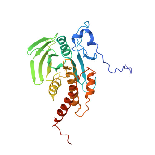
Crystal structure of the catalytic domain of protein-tyrosine phosphatase SHP-1.

Yang, J.Liang, X.Niu, T.Meng, W.Zhao, Z.Zhou, G.W.

(1998) J Biol Chem 273: 28199-28207

  • DOI: https://doi.org/10.1074/jbc.273.43.28199
  • Primary Citation of Related Structures:  
    1GWZ

  • PubMed Abstract: 

    The crystal structures of the protein-tyrosine phosphatase SHP-1 catalytic domain and the complex it forms with the substrate analogue tungstate have been determined and refined to crystallographic R values of 0.209 at 2.5 A resolution and 0.207 at 2.8 A resolution, respectively. Despite low sequence similarity, the catalytic domain of SHP-1 shows high similarity in secondary and tertiary structures with other protein-tyrosine phosphatases (PTPs). In contrast to the conformational changes observed in the crystal structures of PTP1B and Yersinia PTP, the WPD loop (Trp419-Pro428) in the catalytic domain of SHP-1 moves away from the substrate binding pocket after binding the tungstate ion. Sequence alignment and structural analysis suggest that the residues in the WPD loop, especially the amino acid following Asp421, are critical for the movement of WPD loop on binding substrates and the specific activity of protein-tyrosine phosphatases. Our mutagenesis and kinetic measurements have supported this hypothesis.


  • Organizational Affiliation

    Program in Molecular Medicine, University of Massachusetts Medical Center, Worcester, Massachusetts 01605, USA.


Macromolecules
Find similar proteins by:  (by identity cutoff)  |  3D Structure
Entity ID: 1
MoleculeChains Sequence LengthOrganismDetailsImage
SHP-1299Homo sapiensMutation(s): 0 
EC: 3.1.3.48
UniProt & NIH Common Fund Data Resources
Find proteins for P29350 (Homo sapiens)
Explore P29350 
Go to UniProtKB:  P29350
PHAROS:  P29350
GTEx:  ENSG00000111679 
Entity Groups  
Sequence Clusters30% Identity50% Identity70% Identity90% Identity95% Identity100% Identity
UniProt GroupP29350
Sequence Annotations
Expand
  • Reference Sequence
Experimental Data & Validation

Experimental Data

  • Method: X-RAY DIFFRACTION
  • Resolution: 2.50 Å
  • R-Value Free: 0.293 
  • R-Value Work: 0.209 
  • R-Value Observed: 0.209 
  • Space Group: P 1 21 1
Unit Cell:
Length ( Å )Angle ( ˚ )
a = 41.82α = 90
b = 87.78β = 117.4
c = 43γ = 90
Software Package:
Software NamePurpose
AMoREphasing
X-PLORrefinement
DENZOdata reduction
SCALEPACKdata scaling

Structure Validation

View Full Validation Report


Entry History 

Deposition Data

Revision History  (Full details and data files)

  • Version 1.0: 1999-08-22
    Type: Initial release
  • Version 1.1: 2008-03-24
    Changes: Version format compliance
  • Version 1.2: 2011-07-13
    Changes: Version format compliance
  • Version 1.3: 2024-02-07
    Changes: Data collection, Database references
  • Version 1.4: 2024-04-03
    Changes: Refinement description