1GVU

Endothiapepsin complex with H189


Experimental Data Snapshot

  • Method: X-RAY DIFFRACTION
  • Resolution: 0.94 Å
  • R-Value Free: 0.139 
  • R-Value Observed: 0.116 

wwPDB Validation   3D Report Full Report


This is version 2.0 of the entry. See complete history


Literature

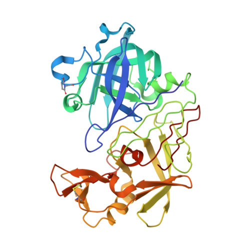
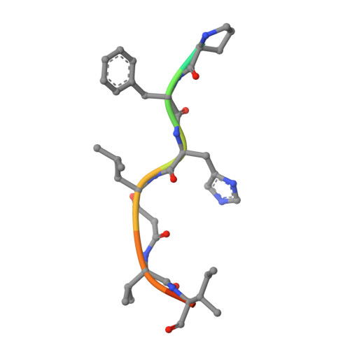
Five Atomic Resolution Structures of Endothiapepsin Inhibitor Complexes: Implications for the Aspartic Proteinase Mechanism

Coates, L.Erskine, P.T.Crump, M.P.Wood, S.P.Cooper, J.B.

(2002) J Mol Biol 318: 1405

  • DOI: https://doi.org/10.1016/s0022-2836(02)00197-3
  • Primary Citation of Related Structures:  
    1GVT, 1GVU, 1GVV, 1GVW, 1GVX

  • PubMed Abstract: 

    Endothiapepsin is derived from the fungus Endothia parasitica and is a member of the aspartic proteinase class of enzymes. This class of enzyme is comprised of two structurally similar lobes, each lobe contributing an aspartic acid residue to form a catalytic dyad that acts to cleave the substrate peptide bond. The three-dimensional structures of endothiapepsin bound to five transition state analogue inhibitors (H189, H256, CP-80,794, PD-129,541 and PD-130,328) have been solved at atomic resolution allowing full anisotropic modelling of each complex. The active sites of the five structures have been studied with a view to studying the catalytic mechanism of the aspartic proteinases by locating the active site protons by carboxyl bond length differences and electron density analysis. In the CP-80,794 structure there is excellent electron density for the hydrogen on the inhibitory statine hydroxyl group which forms a hydrogen bond with the inner oxygen of Asp32. The location of this proton has implications for the catalytic mechanism of the aspartic proteinases as it is consistent with the proposed mechanism in which Asp32 is the negatively charged aspartate. A number of short hydrogen bonds (approximately 2.6 A) with ESD values of around 0.01 A that may have a role in catalysis have been identified within the active site of each structure; the lengths of these bonds have been confirmed using NMR techniques. The possibility and implications of low barrier hydrogen bonds in the active site are considered.


  • Organizational Affiliation

    School of Biological Sciences, University of Southampton, UK. leightonc@bigfoot.com


Macromolecules
Find similar proteins by:  (by identity cutoff)  |  3D Structure
Entity ID: 1
MoleculeChains Sequence LengthOrganismDetailsImage
ENDOTHIAPEPSIN329Cryphonectria parasiticaMutation(s): 0 
EC: 3.4.23.22
UniProt
Find proteins for P11838 (Cryphonectria parasitica)
Explore P11838 
Go to UniProtKB:  P11838
Entity Groups  
Sequence Clusters30% Identity50% Identity70% Identity90% Identity95% Identity100% Identity
UniProt GroupP11838
Sequence Annotations
Expand
  • Reference Sequence

Find similar proteins by:  Sequence   |   3D Structure  

Entity ID: 2
MoleculeChains Sequence LengthOrganismDetailsImage
INHIBITOR, H189B [auth I]10synthetic constructMutation(s): 0 
UniProt
Find proteins for P01017 (Bos taurus)
Explore P01017 
Go to UniProtKB:  P01017
Entity Groups  
Sequence Clusters30% Identity50% Identity70% Identity90% Identity95% Identity100% Identity
UniProt GroupP01017
Sequence Annotations
Expand
  • Reference Sequence
Small Molecules
Ligands 1 Unique
IDChains Name / Formula / InChI Key2D Diagram3D Interactions
SO4
Query on SO4

Download Ideal Coordinates CCD File 
C [auth A],
D [auth A]
SULFATE ION
O4 S
QAOWNCQODCNURD-UHFFFAOYSA-L
Modified Residues  1 Unique
IDChains TypeFormula2D DiagramParent
SUI
Query on SUI
A
L-PEPTIDE LINKINGC6 H8 N2 O4ASP, GLY
Biologically Interesting Molecules (External Reference) 1 Unique
Entity ID: 2
IDChains NameType/Class2D Diagram3D Interactions
PRD_000430
Query on PRD_000430
B [auth I]H 189Peptide-like / Inhibitor
Experimental Data & Validation

Experimental Data

  • Method: X-RAY DIFFRACTION
  • Resolution: 0.94 Å
  • R-Value Free: 0.139 
  • R-Value Observed: 0.116 
  • Space Group: P 1 21 1
Unit Cell:
Length ( Å )Angle ( ˚ )
a = 42.477α = 90
b = 75.779β = 95.43
c = 42.987γ = 90
Software Package:
Software NamePurpose
SHELXL-97refinement
MOSFLMdata reduction
SCALAdata scaling

Structure Validation

View Full Validation Report


Entry History 

Deposition Data

Revision History  (Full details and data files)

  • Version 1.0: 2002-07-04
    Type: Initial release
  • Version 1.1: 2011-07-13
    Changes: Atomic model, Database references, Derived calculations, Non-polymer description, Structure summary, Version format compliance
  • Version 1.2: 2013-03-06
    Changes: Other
  • Version 2.0: 2023-11-15
    Changes: Atomic model, Data collection, Database references, Derived calculations, Source and taxonomy