1GVK

Porcine pancreatic elastase acyl enzyme at 0.95 A resolution


Experimental Data Snapshot

  • Method: X-RAY DIFFRACTION
  • Resolution: 0.94 Å
  • R-Value Free: 0.147 
  • R-Value Observed: 0.122 

wwPDB Validation   3D Report Full Report


This is version 1.7 of the entry. See complete history


Literature

X-Ray Structure of a Serine Protease Acyl-Enzyme Complex at 0.95-A Resolution.

Katona, G.Wilmouth, R.C.Wright, P.A.Berglund, G.I.Hajdu, J.Neutze, R.Schofield, C.J.

(2002) J Biol Chem 277: 21962

  • DOI: https://doi.org/10.1074/jbc.M200676200
  • Primary Citation of Related Structures:  
    1GVK

  • PubMed Abstract: 

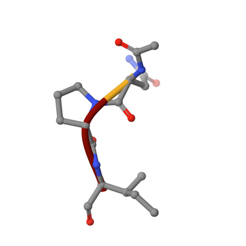
    Kinetic analyses led to the discovery that N-acetylated tripeptides with polar residues at P3 are inhibitors of porcine pancreatic elastase (PPE) that form unusually stable acyl-enzyme complexes. Peptides terminating in a C-terminal carboxylate were more potent than those terminating in a C-terminal amide, suggesting recognition by the oxy-anion hole is important in binding. X-ray diffraction data were recorded to 0.95-A resolution for an acyl-enzyme complex formed between PPE and N-acetyl-Asn-Pro-Ile-CO2H at approximately pH 5. The accuracy of the crystallographic coordinates allows structural issues concerning the mechanism of serine proteases to be addressed. Significantly, the ester bond of the acyl-enzyme showed a high level of planarity, suggesting geometric strain of the ester link is not important during catalysis. Several hydrogen atoms could be clearly identified and were included within the model. In keeping with a recent x-ray structure of subtilisin at 0.78 A (1), limited electron density is visible consistent with the putative location of a hydrogen atom approximately equidistant between the histidine and aspartate residues of the catalytic triad. Comparison of this high resolution crystal structure of the acyl-enzyme complex with that of native elastase at 1.1 A (2) showed that binding of the N-terminal part of the substrate can be accommodated with negligible structural rearrangements. In contrast, comparison with structures obtained as part of "time-resolved" studies on the reacting acyl-enzyme complex at >pH 7 (3) indicate small but significant structural differences, consistent with the proposed synchronization of ester hydrolysis and substrate release.


  • Organizational Affiliation

    Department of Molecular Biotechnology, Chalmers University of Technology, Box 462, 40530 Gothenburg, Sweden.


Macromolecules

Find similar proteins by:  Sequence   |   3D Structure  

Entity ID: 1
MoleculeChains Sequence LengthOrganismDetailsImage
PEPTIDE INHIBITOR4synthetic constructMutation(s): 0 
Sequence Annotations
Expand
  • Reference Sequence
Find similar proteins by:  (by identity cutoff)  |  3D Structure
Entity ID: 2
MoleculeChains Sequence LengthOrganismDetailsImage
ELASTASE 1240Sus scrofaMutation(s): 0 
EC: 3.4.21.36
UniProt
Find proteins for P00772 (Sus scrofa)
Explore P00772 
Go to UniProtKB:  P00772
Entity Groups  
Sequence Clusters30% Identity50% Identity70% Identity90% Identity95% Identity100% Identity
UniProt GroupP00772
Sequence Annotations
Expand
  • Reference Sequence
Experimental Data & Validation

Experimental Data

  • Method: X-RAY DIFFRACTION
  • Resolution: 0.94 Å
  • R-Value Free: 0.147 
  • R-Value Observed: 0.122 
  • Space Group: P 21 21 21
Unit Cell:
Length ( Å )Angle ( ˚ )
a = 50.176α = 90
b = 57.778β = 90
c = 74.409γ = 90
Software Package:
Software NamePurpose
SHELXL-97refinement
MOSFLMdata reduction
SCALAdata scaling
CNSphasing

Structure Validation

View Full Validation Report


Entry History 

Deposition Data

Revision History  (Full details and data files)

  • Version 1.0: 2002-07-19
    Type: Initial release
  • Version 1.1: 2011-07-13
    Changes: Atomic model, Database references, Derived calculations, Non-polymer description, Structure summary, Version format compliance
  • Version 1.2: 2015-04-15
    Changes: Atomic model, Database references, Derived calculations, Other
  • Version 1.3: 2015-09-16
    Changes: Source and taxonomy
  • Version 1.4: 2018-06-20
    Changes: Advisory, Data collection, Derived calculations
  • Version 1.5: 2019-05-08
    Changes: Advisory, Data collection, Derived calculations, Experimental preparation
  • Version 1.6: 2019-05-22
    Changes: Data collection, Refinement description
  • Version 1.7: 2023-12-13
    Changes: Advisory, Data collection, Database references, Derived calculations, Other, Refinement description