1GV3

The 2.0 Angstrom resolution structure of the catalytic portion of a cyanobacterial membrane-bound manganese superoxide dismutase


Experimental Data Snapshot

  • Method: X-RAY DIFFRACTION
  • Resolution: 2.00 Å
  • R-Value Free: 0.211 
  • R-Value Work: 0.188 
  • R-Value Observed: 0.188 

wwPDB Validation   3D Report Full Report


This is version 1.5 of the entry. See complete history


Literature

The 2.0A Resolution Structure of the Catalytic Portion of a Cyanobacterial Membrane-Bound Manganese Superoxide Dismutase

Atzenhofer, W.Regelsberger, G.Jacob, U.Peschek, G.A.Furtmuller, P.Huber, R.Obinger, C.

(2002) J Mol Biol 321: 479

  • DOI: https://doi.org/10.1016/s0022-2836(02)00624-1
  • Primary Citation of Related Structures:  
    1GV3

  • PubMed Abstract: 

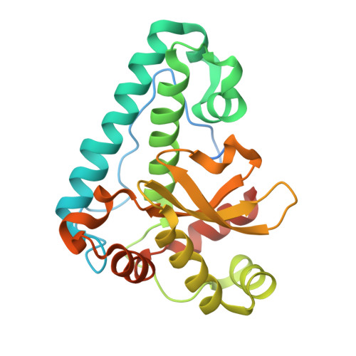
    Cyanobacteria are shown to be unique in containing membrane-bound manganese superoxide dismutases (MnSOD). They are homodimeric type 2 membrane proteins that protect this phototrophic organism against oxidative stress. We have determined, for the first time, the 2.0A resolution structure of the catalytic portion of the MnSOD from the filamentous cyanobacterium Anabaena PCC 7120. Within each subunit, both the N-terminal helical hairpin (His94 and His145) and the C-terminal alpha/beta domain (His232 and Asp228) contribute ligands to the catalytic manganese site. Together with a water or hydroxide ion (OH(x)) a five-coordinated trigonal bipyramidal geometry is formed, with OH(x) and His90 forming the axial ligands and manganese shifted out of the equatorial plane in the direction of OH(x). The ligands including OH(x) are tightly constrained by hydrogen bonding with surrounding residues either from the same monomer (Tyr98, Asn144, Trp194, Gln213, Val229, Trp230) or from the neighbouring subunit (Glu231, Tyr235). This underlines the important role of the symmetric dimeric structure of MnSODs in contributing elements to both the active site and the substrate funnel. The Mn cdots, three dots, centered Mn distance (18.4A) is bridged by the hydrogen-bonded His232 of one monomer with Glu231 of the other monomer. A detailed discussion of the structure, a comparison with known structures of soluble MnSODs as well as a model of the cyanobacterial membrane-bound MnSOD is presented.


  • Organizational Affiliation

    Max-Planck-Institut für Biochemie/Abt. Strukturforschung, Am Klopferspitz 18a, D-82152 Martinsried/Planegg, Munich, Germany.


Macromolecules
Find similar proteins by:  (by identity cutoff)  |  3D Structure
Entity ID: 1
MoleculeChains Sequence LengthOrganismDetailsImage
MANGANESE SUPEROXIDE DISMUTASE
A, B
248Nostoc sp. PCC 7120 = FACHB-418Mutation(s): 0 
UniProt
Find proteins for Q8Z0M1 (Nostoc sp. (strain PCC 7120 / SAG 25.82 / UTEX 2576))
Explore Q8Z0M1 
Go to UniProtKB:  Q8Z0M1
Entity Groups  
Sequence Clusters30% Identity50% Identity70% Identity90% Identity95% Identity100% Identity
UniProt GroupQ8Z0M1
Sequence Annotations
Expand
  • Reference Sequence
Small Molecules
Ligands 1 Unique
IDChains Name / Formula / InChI Key2D Diagram3D Interactions
MN
Query on MN

Download Ideal Coordinates CCD File 
C [auth A],
D [auth B]
MANGANESE (II) ION
Mn
WAEMQWOKJMHJLA-UHFFFAOYSA-N
Experimental Data & Validation

Experimental Data

  • Method: X-RAY DIFFRACTION
  • Resolution: 2.00 Å
  • R-Value Free: 0.211 
  • R-Value Work: 0.188 
  • R-Value Observed: 0.188 
  • Space Group: H 3
Unit Cell:
Length ( Å )Angle ( ˚ )
a = 151.32α = 90
b = 151.32β = 90
c = 69.28γ = 120
Software Package:
Software NamePurpose
CNSrefinement
MOSFLMdata reduction
AMoREphasing

Structure Validation

View Full Validation Report


Entry History 

Deposition Data

Revision History  (Full details and data files)

  • Version 1.0: 2002-08-08
    Type: Initial release
  • Version 1.1: 2011-06-02
    Changes: Version format compliance
  • Version 1.2: 2011-07-13
    Changes: Version format compliance
  • Version 1.3: 2018-04-04
    Changes: Data collection
  • Version 1.4: 2018-05-23
    Changes: Data collection
  • Version 1.5: 2023-12-13
    Changes: Data collection, Database references, Derived calculations, Refinement description