1GUH

Structure determination and refinement of human alpha class glutathione transferase A1-1, and a comparison with the MU and PI class enzymes


Experimental Data Snapshot

  • Method: X-RAY DIFFRACTION
  • Resolution: 2.60 Å
  • R-Value Work: 0.229 
  • R-Value Observed: 0.229 

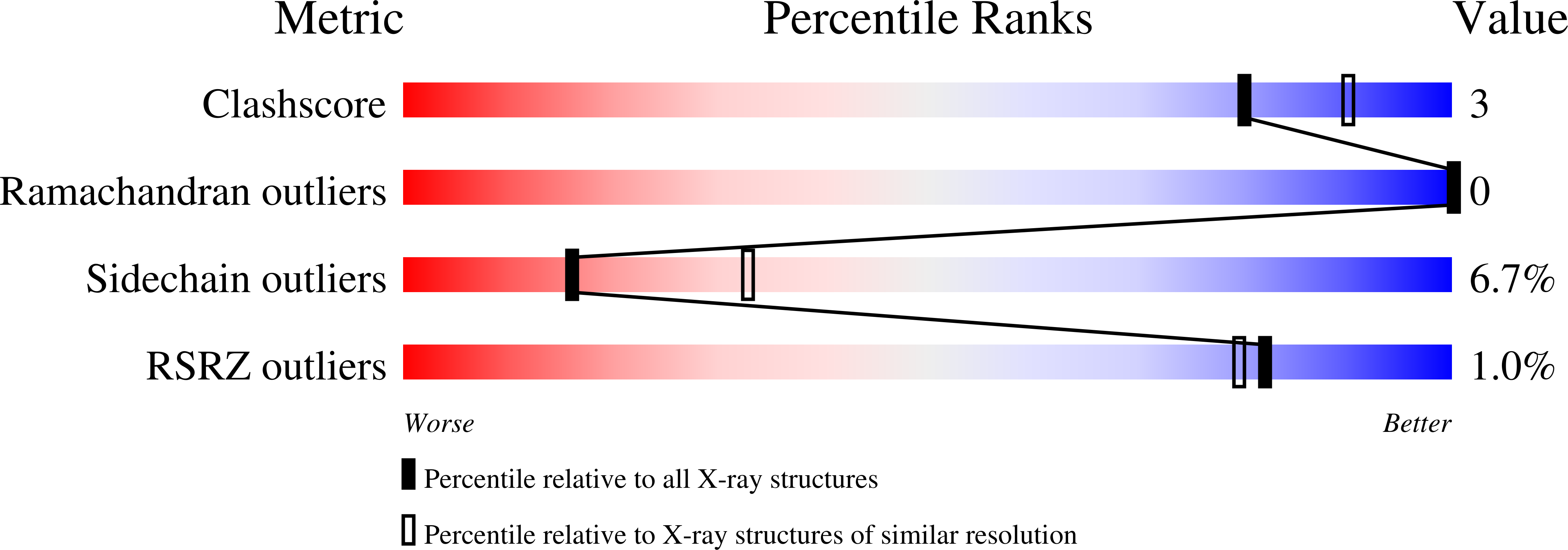
wwPDB Validation   3D Report Full Report


Ligand Structure Quality Assessment 


This is version 1.3 of the entry. See complete history


Literature

Structure determination and refinement of human alpha class glutathione transferase A1-1, and a comparison with the Mu and Pi class enzymes.

Sinning, I.Kleywegt, G.J.Cowan, S.W.Reinemer, P.Dirr, H.W.Huber, R.Gilliland, G.L.Armstrong, R.N.Ji, X.Board, P.G.Olin, B.Mannervik, B.Jones, T.A.

(1993) J Mol Biol 232: 192-212

  • DOI: https://doi.org/10.1006/jmbi.1993.1376
  • Primary Citation of Related Structures:  
    1GUH

  • PubMed Abstract: 

    The crystal structure of human alpha class glutathione transferase A1-1 has been determined and refined to a resolution of 2.6 A. There are two copies of the dimeric enzyme in the asymmetric unit. Each monomer is built from two domains. A bound inhibitor, S-benzyl-glutathione, is primarily associated with one of these domains via a network of hydrogen bonds and salt-links. In particular, the sulphur atom of the inhibitor forms a hydrogen bond to the hydroxyl group of Tyr9 and the guanido group of Arg15. The benzyl group of the inhibitor is completely buried in a hydrophobic pocket. The structure shows an overall similarity to the mu and pi class enzymes particularly in the glutathione-binding domain". The main difference concerns the extended C terminus of the alpha class enzyme which forms an extra alpha-helix that blocks one entrance to the active site and makes up part of the substrate binding site.


  • Organizational Affiliation

    Department of Molecular Biology, Uppsala University Biomedical Center, Sweden.


Macromolecules
Find similar proteins by:  (by identity cutoff)  |  3D Structure
Entity ID: 1
MoleculeChains Sequence LengthOrganismDetailsImage
GLUTATHIONE S-TRANSFERASE A1-1
A, B, C, D
221Homo sapiensMutation(s): 0 
EC: 2.5.1.18
UniProt & NIH Common Fund Data Resources
Find proteins for P08263 (Homo sapiens)
Explore P08263 
Go to UniProtKB:  P08263
PHAROS:  P08263
GTEx:  ENSG00000243955 
Entity Groups  
Sequence Clusters30% Identity50% Identity70% Identity90% Identity95% Identity100% Identity
UniProt GroupP08263
Sequence Annotations
Expand
  • Reference Sequence
Experimental Data & Validation

Experimental Data

  • Method: X-RAY DIFFRACTION
  • Resolution: 2.60 Å
  • R-Value Work: 0.229 
  • R-Value Observed: 0.229 
  • Space Group: C 1 2 1
Unit Cell:
Length ( Å )Angle ( ˚ )
a = 100.8α = 90
b = 95.4β = 92.4
c = 105.2γ = 90
Software Package:
Software NamePurpose
X-PLORmodel building
X-PLORrefinement
X-PLORphasing

Structure Validation

View Full Validation Report


Ligand Structure Quality Assessment 


Entry History 

Deposition Data

Revision History  (Full details and data files)

  • Version 1.0: 1993-10-31
    Type: Initial release
  • Version 1.1: 2008-03-24
    Changes: Version format compliance
  • Version 1.2: 2011-07-13
    Changes: Version format compliance
  • Version 1.3: 2024-02-07
    Changes: Data collection, Database references, Derived calculations, Other