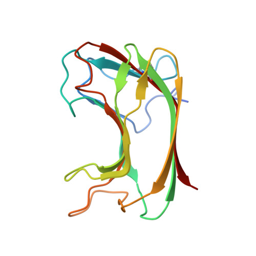
1GU3

CBM4 structure and function


Experimental Data Snapshot

  • Method: X-RAY DIFFRACTION
  • Resolution: 2.30 Å
  • R-Value Free: 0.257 
  • R-Value Work: 0.209 
  • R-Value Observed: 0.217 

wwPDB Validation   3D Report Full Report


This is version 2.1 of the entry. See complete history


Literature

Differential Oligosaccharide Recognition by Evolutionarily-Related Beta-1,4 and Beta-1,3 Glucan-Binding Modules

Boraston, A.B.Nurizzo, D.Notenboom, V.Ducros, V.Rose, D.R.Kilburn, D.G.Davies, G.J.

(2002) J Mol Biol 319: 1143

  • DOI: https://doi.org/10.1016/S0022-2836(02)00374-1
  • Primary Citation of Related Structures:  
    1GU3, 1GUI

  • PubMed Abstract: 

    Enzymes active on complex carbohydrate polymers frequently have modular structures in which a catalytic domain is appended to one or more carbohydrate-binding modules (CBMs). Although CBMs have been classified into a number of families based upon sequence, many closely related CBMs are specific for different polysaccharides. In order to provide a structural rationale for the recognition of different polysaccharides by CBMs displaying a conserved fold, we have studied the thermodynamics of binding and three-dimensional structures of the related family 4 CBMs from Cellulomonas fimi Cel9B and Thermotoga maritima Lam16A in complex with their ligands, beta-1,4 and beta-1,3 linked gluco-oligosaccharides, respectively. These two CBMs use a structurally conserved constellation of aromatic and polar amino acid side-chains that interact with sugars in two of the five binding subsites. Differences in the length and conformation of loops in non-conserved regions create binding-site topographies that complement the known solution conformations of their respective ligands. Thermodynamics interpreted in the light of structural information highlights the differential role of water in the interaction of these CBMs with their respective oligosaccharide ligands.


  • Organizational Affiliation

    Protein Engineering Network of Centres of Excellence, Edmonton, Alberta, Canada T6G 2S2.


Macromolecules
Find similar proteins by:  (by identity cutoff)  |  3D Structure
Entity ID: 1
MoleculeChains Sequence LengthOrganismDetailsImage
ENDOGLUCANASE C149Cellulomonas fimiMutation(s): 0 
EC: 3.2.1.4
UniProt
Find proteins for P14090 (Cellulomonas fimi (strain ATCC 484 / DSM 20113 / JCM 1341 / NBRC 15513 / NCIMB 8980 / NCTC 7547))
Explore P14090 
Go to UniProtKB:  P14090
Entity Groups  
Sequence Clusters30% Identity50% Identity70% Identity90% Identity95% Identity100% Identity
UniProt GroupP14090
Sequence Annotations
Expand
  • Reference Sequence
Oligosaccharides

Help

Entity ID: 2
MoleculeChains Length2D Diagram Glycosylation3D Interactions
beta-D-glucopyranose-(1-4)-beta-D-glucopyranose-(1-4)-beta-D-glucopyranose-(1-4)-beta-D-glucopyranose-(1-4)-beta-D-glucopyranose
B
5N/A
Glycosylation Resources
GlyTouCan:  G91683DU
GlyCosmos:  G91683DU
Biologically Interesting Molecules (External Reference) 1 Unique
Experimental Data & Validation

Experimental Data

  • Method: X-RAY DIFFRACTION
  • Resolution: 2.30 Å
  • R-Value Free: 0.257 
  • R-Value Work: 0.209 
  • R-Value Observed: 0.217 
  • Space Group: P 31 2 1
Unit Cell:
Length ( Å )Angle ( ˚ )
a = 77.634α = 90
b = 77.634β = 90
c = 55.63γ = 120
Software Package:
Software NamePurpose
REFMACrefinement
DENZOdata reduction
SCALEPACKdata scaling
AMoREphasing

Structure Validation

View Full Validation Report


Entry History 

Deposition Data

Revision History  (Full details and data files)

  • Version 1.0: 2002-09-26
    Type: Initial release
  • Version 1.1: 2011-05-08
    Changes: Version format compliance
  • Version 1.2: 2011-07-13
    Changes: Version format compliance
  • Version 2.0: 2020-07-29
    Type: Remediation
    Reason: Carbohydrate remediation
    Changes: Atomic model, Data collection, Derived calculations, Other, Structure summary
  • Version 2.1: 2023-12-13
    Changes: Data collection, Database references, Refinement description, Structure summary