


Time-Resolved and Static-Ensemble Structural Chemistry of Hydroxymethylbilane Synthase
Helliwell, J.R., Nieh, Y.P., Habash, J., Faulder, P.F., Raftery, J., Cianci, M., Wulff, M., Hadener, A.(2003) Faraday Discuss 122: 131
- PubMed: 12555854
- DOI: https://doi.org/10.1039/b201331b
- Primary Citation of Related Structures:
1GTK - PubMed Abstract:
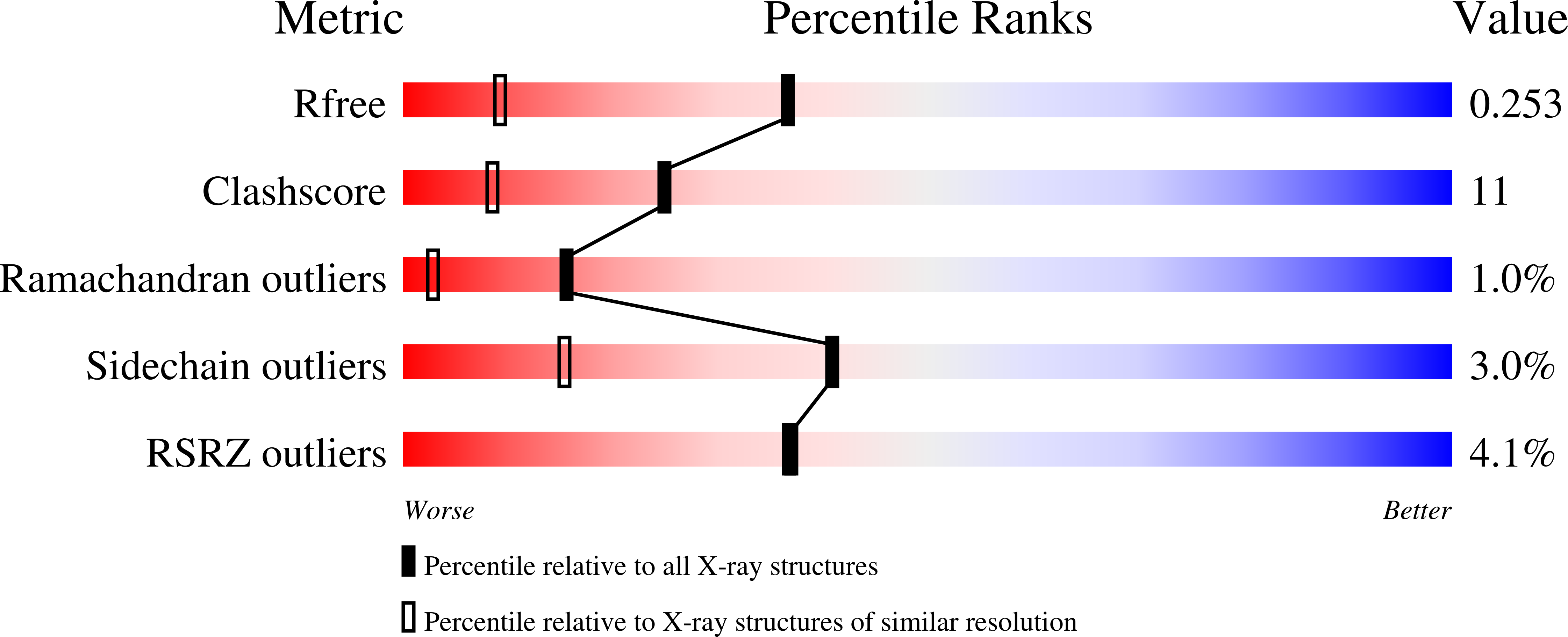
The enzyme hydroxymethylbilane synthase (HMBS, EC 4.3.1.8), 313 amino acid residues and MW 34 kDa, also known as porphobilinogen deaminase (PBGD), catalyses the stepwise polymerization of four molecules of porphobilinogen (PBG) to the linear tetrapyrrole 1-hydroxymethylbilane. Several crystallographic structures of HMBS have been previously determined, most recently including by time-resolved Laue protein crystallography of the Lys59Gln mutant form with reaction initiation undertaken by use of a flow cell carrying the substrate PBG. In this paper we review these structures and add new molecular graphics representations and analyses. Moreover we present a new structure refined at 1.66 A resolution using diffraction data recorded at cryo-temperature (100 K) in an attempt at trapping the polypeptide loop (residues 47 to 58) in the vicinity of the enzyme active site, missing in all previous structure determinations. This loop still has not appeared in the electron density maps, in spite of the advantage of cryo-temperature, but nevertheless the 1.66 A cryo-structure extends the ensemble of known HMBS structures. The cryomodel of protein, cofactor and 320 bound water molecules has been refined to a final R-factor and R-free of 0.198 and 0.247 respectively; the PDB deposition codes, coordinates and structure factors are 1GTK and R1GTKSF respectively. Finally a protein comparison study is presented of the Mycobacterium tuberculosis (MTb) HMBS, with the E. coli HMBS. This has been done as preparation for future structural studies on the MTb HMBS from this important disease bearing organism. The overall amino acid sequence identity is 41%. Most interestingly there is a two-residue reduction in length of the loop referred to above (Asp 50 and Gly 58 being missing in the MTb form). This gives the hope that this loop will be less flexible and thus might become visible to crystallographic analysis.
Organizational Affiliation:
Laboratory of Structural Chemistry, Department of Chemistry, University of Manchester, Manchester, UK M13 9PL. j.r.helliwell@dl.ac.uk


