1GSY

GLUTATHIONE S-TRANSFERASE YFYF, CLASS PI, COMPLEXED WITH GLUTATHIONE


Experimental Data Snapshot

  • Method: X-RAY DIFFRACTION
  • Resolution: 2.44 Å
  • R-Value Work: 0.194 
  • R-Value Observed: 0.194 

wwPDB Validation   3D Report Full Report


Ligand Structure Quality Assessment 


This is version 1.4 of the entry. See complete history


Literature

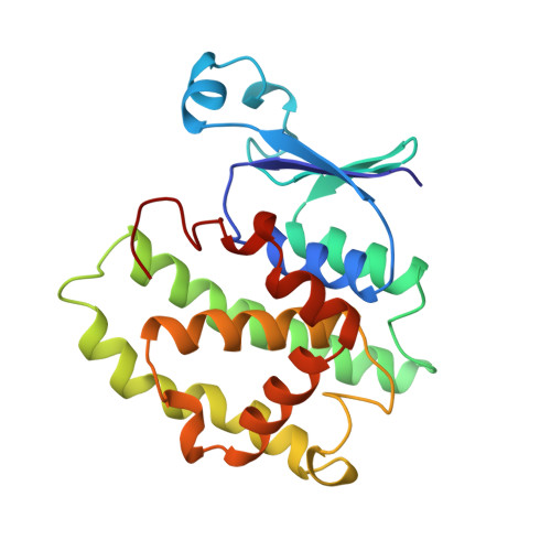
The three-dimensional structure of a class-Pi glutathione S-transferase complexed with glutathione: the active-site hydration provides insights into the reaction mechanism.

Parraga, A.Garcia-Saez, I.Walsh, S.B.Mantle, T.J.Coll, M.

(1998) Biochem J 333 ( Pt 3): 811-816

  • DOI: https://doi.org/10.1042/bj3330811
  • Primary Citation of Related Structures:  
    1GSY

  • PubMed Abstract: 

    The structure of mouse liver glutathione S-transferase P1-1 complexed with its substrate glutathione (GSH) has been determined by X-ray diffraction analysis. No conformational changes in the glutathione moiety or in the protein, other than small adjustments of some side chains, are observed when compared with glutathione adduct complexes. Our structure confirms that the role of Tyr-7 is to stabilize the thiolate by hydrogen bonding and to position it in the right orientation. A comparison of the enzyme-GSH structure reported here with previously described structures reveals rearrangements in a well-defined network of water molecules in the active site. One of these water molecules (W0), identified in the unliganded enzyme (carboxymethylated at Cys-47), is displaced by the binding of GSH, and a further water molecule (W4) is displaced following the binding of the electrophilic substrate and the formation of the glutathione conjugate. The possibility that one of these water molecules participates in the proton abstraction from the glutathione thiol is discussed.


  • Organizational Affiliation

    Departament de Biologia Molecular i Cel.lular, Centre d'Investigació i Desenvolupament-CSIC, Jordi Girona 18-26, 08034 Barcelona, Spain.


Macromolecules
Find similar proteins by:  (by identity cutoff)  |  3D Structure
Entity ID: 1
MoleculeChains Sequence LengthOrganismDetailsImage
GLUTATHIONE S-TRANSFERASE CLASS PI
A, B
209Mus musculusMutation(s): 0 
EC: 2.5.1.18
UniProt
Find proteins for P19157 (Mus musculus)
Explore P19157 
Go to UniProtKB:  P19157
Entity Groups  
Sequence Clusters30% Identity50% Identity70% Identity90% Identity95% Identity100% Identity
UniProt GroupP19157
Sequence Annotations
Expand
  • Reference Sequence
Experimental Data & Validation

Experimental Data

  • Method: X-RAY DIFFRACTION
  • Resolution: 2.44 Å
  • R-Value Work: 0.194 
  • R-Value Observed: 0.194 
  • Space Group: C 2 2 21
Unit Cell:
Length ( Å )Angle ( ˚ )
a = 142.68α = 90
b = 162.57β = 90
c = 63.32γ = 90
Software Package:
Software NamePurpose
X-PLORmodel building
X-PLORrefinement
XDSdata reduction
X-PLORphasing

Structure Validation

View Full Validation Report


Ligand Structure Quality Assessment 


Entry History 

Deposition Data

Revision History  (Full details and data files)

  • Version 1.0: 1997-11-19
    Type: Initial release
  • Version 1.1: 2008-03-10
    Changes: Version format compliance
  • Version 1.2: 2011-07-13
    Changes: Version format compliance
  • Version 1.3: 2011-12-14
    Changes: Non-polymer description
  • Version 1.4: 2023-08-09
    Changes: Database references, Derived calculations, Other, Refinement description