1GQM

The structure of S100A12 in a hexameric form and its proposed role in receptor signalling


Experimental Data Snapshot

  • Method: X-RAY DIFFRACTION
  • Resolution: 2.70 Å
  • R-Value Free: 0.281 
  • R-Value Work: 0.217 
  • R-Value Observed: 0.219 

wwPDB Validation   3D Report Full Report


This is version 1.4 of the entry. See complete history


Literature

The Structure of S100A12 in a Hexameric Form and its Proposed Role in Receptor Signalling

Moroz, O.V.Antson, A.A.Dodson, E.J.Burrell, H.J.Grist, S.J.Lloyd, R.M.Maitland, N.J.Dodson, G.G.Wilson, K.S.Lukanidin, E.Bronstein, I.B.

(2002) Acta Crystallogr D Biol Crystallogr 58: 407

  • DOI: https://doi.org/10.1107/s0907444901021278
  • Primary Citation of Related Structures:  
    1GQM

  • PubMed Abstract: 

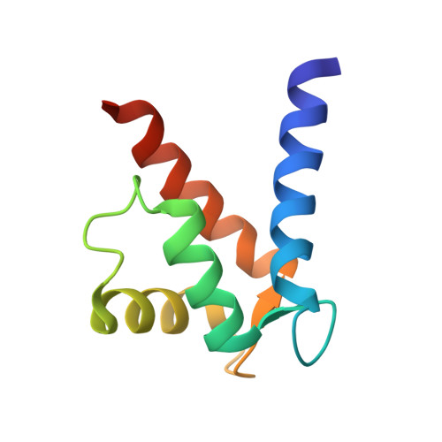
    S100A12 is a member of the S100 subfamily of EF-hand calcium-binding proteins; it has been shown to be one of the ligands of the 'receptor for advanced glycation end products' (RAGE) that belongs to the immunoglobulin superfamily and is involved in diabetes, Alzheimer's disease, inflammation and tumour invasion. The structure of the dimeric form of native S100A12 from human granulocytes in the presence of calcium in space group R3 has previously been reported. Here, the structure of a second crystal form in space group P2(1) (unit-cell parameters a = 53.9, b = 100.5, c = 112.7A, beta = 94.6 degrees) solved at 2.7A resolution by molecular replacement using the R3 structure as a search model is reported. Like most S100 proteins, S100A12 is a dimer. However, in the P2(1) crystal form dimers of S100A12 are arranged in a spherical hexameric assembly with an external diameter of about 55 A stabilized by calcium ions bound between adjacent dimers. The putative target-binding sites of S100A12 are located at the outer surface of the hexamer, making it possible for the hexamer to bind several targets. It is proposed that the S100A12 hexameric assembly might interact with three extracellular domains of the receptor, bringing them together into large trimeric assemblies.


  • Organizational Affiliation

    Department of Chemistry, University of York, York YO10 5DD, England.


Macromolecules
Find similar proteins by:  (by identity cutoff)  |  3D Structure
Entity ID: 1
MoleculeChains Sequence LengthOrganismDetailsImage
CALGRANULIN C
A, B, C, D, E
A, B, C, D, E, F, G, H, I, J, K, L
91Homo sapiensMutation(s): 0 
UniProt & NIH Common Fund Data Resources
Find proteins for P80511 (Homo sapiens)
Explore P80511 
Go to UniProtKB:  P80511
PHAROS:  P80511
GTEx:  ENSG00000163221 
Entity Groups  
Sequence Clusters30% Identity50% Identity70% Identity90% Identity95% Identity100% Identity
UniProt GroupP80511
Sequence Annotations
Expand
  • Reference Sequence
Small Molecules
Ligands 1 Unique
IDChains Name / Formula / InChI Key2D Diagram3D Interactions
CA
Query on CA

Download Ideal Coordinates CCD File 
AA [auth E]
BA [auth E]
CA [auth F]
DA [auth F]
EA [auth G]
AA [auth E],
BA [auth E],
CA [auth F],
DA [auth F],
EA [auth G],
FA [auth G],
GA [auth G],
HA [auth H],
IA [auth H],
JA [auth H],
KA [auth I],
LA [auth I],
M [auth A],
MA [auth J],
N [auth A],
NA [auth J],
O [auth A],
OA [auth J],
P [auth B],
PA [auth J],
Q [auth B],
QA [auth K],
R [auth B],
RA [auth K],
S [auth C],
SA [auth K],
T [auth C],
TA [auth K],
U [auth C],
UA [auth L],
V [auth C],
VA [auth L],
W [auth D],
X [auth D],
Y [auth E],
Z [auth E]
CALCIUM ION
Ca
BHPQYMZQTOCNFJ-UHFFFAOYSA-N
Experimental Data & Validation

Experimental Data

  • Method: X-RAY DIFFRACTION
  • Resolution: 2.70 Å
  • R-Value Free: 0.281 
  • R-Value Work: 0.217 
  • R-Value Observed: 0.219 
  • Space Group: P 1 21 1
Unit Cell:
Length ( Å )Angle ( ˚ )
a = 53.875α = 90
b = 100.501β = 94.55
c = 112.687γ = 90
Software Package:
Software NamePurpose
REFMACrefinement
DENZOdata reduction
SCALEPACKdata scaling
AMoREphasing

Structure Validation

View Full Validation Report


Entry History 

Revision History  (Full details and data files)

  • Version 1.0: 2002-02-28
    Type: Initial release
  • Version 1.1: 2011-07-13
    Changes: Advisory, Version format compliance
  • Version 1.2: 2011-09-28
    Changes: Atomic model, Derived calculations, Non-polymer description
  • Version 1.3: 2019-05-08
    Changes: Advisory, Data collection, Experimental preparation, Other
  • Version 1.4: 2023-12-13
    Changes: Advisory, Data collection, Database references, Derived calculations, Other, Refinement description