1GQH

Quercetin 2,3-dioxygenase in complex with the inhibitor kojic acid


Experimental Data Snapshot

  • Method: X-RAY DIFFRACTION
  • Resolution: 2.15 Å
  • R-Value Free: 0.228 
  • R-Value Work: 0.178 
  • R-Value Observed: 0.181 

wwPDB Validation   3D Report Full Report


Ligand Structure Quality Assessment 


This is version 2.0 of the entry. See complete history


Literature

Functional Analysis of the Copper-Dependent Quercetin 2,3-Dioxygenase.1.Ligand-Induced Coordination Changes Probed by X-Ray Crystallography: Inhibition, Ordering Effect and Mechanistic Insights

Steiner, R.A.Kooter, I.M.Dijkstra, B.W.

(2002) Biochemistry 41: 7955

  • DOI: https://doi.org/10.1021/bi0159736
  • Primary Citation of Related Structures:  
    1GQG, 1GQH

  • PubMed Abstract: 

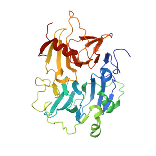
    The crystal structures of the copper-dependent Aspergillus japonicus quercetin 2,3-dioxygenase (2,3QD) complexed with the inhibitors diethyldithiocarbamate (DDC) and kojic acid (KOJ) are reported at 1.70 and 2.15 A resolution, respectively. Both inhibitors asymmetrically chelate the metal center and assume a common orientation in the active site cleft. Their molecular plane blocks access to the inner portion of the cavity which is lined by the side chains of residues Met51, Thr53, Phe75, Phe114, and Met123 and which is believed to bind the flavonol B-ring of the natural substrate. The binding of the inhibitors brings order into the mixed coordination observed in the native enzyme. DDC and KOJ induce a single conformation of the Glu73 side chain, although in different ways. In the presence of DDC, Glu73 is detached from the copper ion with its carboxylate moiety pointing away from the active site cavity. In contrast, when KOJ is bound, Glu73 ligates the Cu ion through its O(epsilon)(1) atom with a monodentate geometry. Compared to the native coordinating conformation, this conformation is approximately 90 degrees rotated about the chi(3) angle. This latter Glu73 conformation is compatible with the presence of a bound substrate.


  • Organizational Affiliation

    Laboratory of Biophysical Chemistry, Department of Chemistry, University of Groningen, Nijenborgh 4, 9747 AG Groningen, The Netherlands.


Macromolecules
Find similar proteins by:  (by identity cutoff)  |  3D Structure
Entity ID: 1
MoleculeChains Sequence LengthOrganismDetailsImage
QUERCETIN 2,3-DIOXYGENASE
A, B, C, D
350Aspergillus japonicusMutation(s): 0 
EC: 1.13.11.24
UniProt
Find proteins for Q7SIC2 (Aspergillus japonicus)
Explore Q7SIC2 
Go to UniProtKB:  Q7SIC2
Entity Groups  
Sequence Clusters30% Identity50% Identity70% Identity90% Identity95% Identity100% Identity
UniProt GroupQ7SIC2
Sequence Annotations
Expand
  • Reference Sequence
Oligosaccharides

Help

Entity ID: 2
MoleculeChains Length2D Diagram Glycosylation3D Interactions
alpha-D-mannopyranose-(1-4)-2-acetamido-2-deoxy-beta-D-glucopyranose-(1-4)-2-acetamido-2-deoxy-beta-D-glucopyranose
E
3N-Glycosylation
Glycosylation Resources
GlyTouCan:  G62182OO
GlyCosmos:  G62182OO
GlyGen:  G62182OO
Entity ID: 3
MoleculeChains Length2D Diagram Glycosylation3D Interactions
alpha-D-mannopyranose-(1-3)-alpha-D-mannopyranose-(1-4)-2-acetamido-2-deoxy-beta-D-glucopyranose-(1-4)-2-acetamido-2-deoxy-beta-D-glucopyranose
F, G
4N-Glycosylation
Glycosylation Resources
GlyTouCan:  G48068RF
GlyCosmos:  G48068RF
GlyGen:  G48068RF
Entity ID: 4
MoleculeChains Length2D Diagram Glycosylation3D Interactions
2-acetamido-2-deoxy-beta-D-glucopyranose-(1-4)-2-acetamido-2-deoxy-beta-D-glucopyranose
H
2N-Glycosylation
Glycosylation Resources
GlyTouCan:  G42666HT
GlyCosmos:  G42666HT
GlyGen:  G42666HT
Small Molecules
Ligands 3 Unique
IDChains Name / Formula / InChI Key2D Diagram3D Interactions
NAG
Query on NAG

Download Ideal Coordinates CCD File 
AA [auth D]
BA [auth D]
K [auth A]
L [auth A]
M [auth A]
AA [auth D],
BA [auth D],
K [auth A],
L [auth A],
M [auth A],
N [auth A],
Q [auth B],
R [auth B],
S [auth B],
V [auth C],
W [auth C],
X [auth C]
2-acetamido-2-deoxy-beta-D-glucopyranose
C8 H15 N O6
OVRNDRQMDRJTHS-FMDGEEDCSA-N
KOJ
Query on KOJ

Download Ideal Coordinates CCD File 
I [auth A],
O [auth B],
T [auth C],
Y [auth D]
5-HYDROXY-2-(HYDROXYMETHYL)-4H-PYRAN-4-ONE
C6 H6 O4
BEJNERDRQOWKJM-UHFFFAOYSA-N
CU
Query on CU

Download Ideal Coordinates CCD File 
J [auth A],
P [auth B],
U [auth C],
Z [auth D]
COPPER (II) ION
Cu
JPVYNHNXODAKFH-UHFFFAOYSA-N
Experimental Data & Validation

Experimental Data

  • Method: X-RAY DIFFRACTION
  • Resolution: 2.15 Å
  • R-Value Free: 0.228 
  • R-Value Work: 0.178 
  • R-Value Observed: 0.181 
  • Space Group: P 1 21 1
Unit Cell:
Length ( Å )Angle ( ˚ )
a = 108.64α = 90
b = 55.396β = 98.26
c = 124.425γ = 90
Software Package:
Software NamePurpose
REFMACrefinement
DENZOdata reduction
SCALEPACKdata scaling

Structure Validation

View Full Validation Report


Ligand Structure Quality Assessment 


Entry History 

Deposition Data

Revision History  (Full details and data files)

  • Version 1.0: 2002-06-21
    Type: Initial release
  • Version 1.1: 2011-07-13
    Changes: Advisory, Version format compliance
  • Version 1.2: 2019-05-08
    Changes: Data collection, Derived calculations, Experimental preparation, Other
  • Version 2.0: 2020-07-29
    Type: Remediation
    Reason: Carbohydrate remediation
    Changes: Advisory, Atomic model, Data collection, Derived calculations, Other, Structure summary