


Glycogen Phosphorylase B: Description of the Protein Structure
Acharya, K.R., Stuart, D.I., Varvill, K.M., Johnson, L.N.(1991) Glycogen Phosphorylase B: Description Of The Protein Structure : 1
Experimental Data Snapshot
wwPDB Validation 3D Report Full Report

(1991) Glycogen Phosphorylase B: Description Of The Protein Structure : 1
Entity ID: 1 | |||||
|---|---|---|---|---|---|
| Molecule | Chains | Sequence Length | Organism | Details | Image |
| GLYCOGEN PHOSPHORYLASE B | 842 | Oryctolagus cuniculus | Mutation(s): 0 EC: 2.4.1.1 | 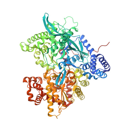 | |
UniProt | |||||
Find proteins for P00489 (Oryctolagus cuniculus) Explore P00489 Go to UniProtKB: P00489 | |||||
Entity Groups | |||||
| Sequence Clusters | 30% Identity50% Identity70% Identity90% Identity95% Identity100% Identity | ||||
| UniProt Group | P00489 | ||||
Sequence AnnotationsExpand | |||||
| |||||
| Ligands 1 Unique | |||||
|---|---|---|---|---|---|
| ID | Chains | Name / Formula / InChI Key | 2D Diagram | 3D Interactions | |
| PLP Query on PLP | B [auth A] | PYRIDOXAL-5'-PHOSPHATE C8 H10 N O6 P NGVDGCNFYWLIFO-UHFFFAOYSA-N |  | ||
| Length ( Å ) | Angle ( ˚ ) |
|---|---|
| a = 128.5 | α = 90 |
| b = 128.5 | β = 90 |
| c = 116.3 | γ = 90 |
| Software Name | Purpose |
|---|---|
| PROLSQ | refinement |
RCSB PDB (citation) is hosted by
RCSB PDB is a member of the

