1GNI

HUMAN SERUM ALBUMIN COMPLEXED WITH cis-9-OCTADECENOIC ACID (OLEIC ACID)


Experimental Data Snapshot

  • Method: X-RAY DIFFRACTION
  • Resolution: 2.40 Å
  • R-Value Free: 0.242 
  • R-Value Work: 0.209 
  • R-Value Observed: 0.209 

wwPDB Validation   3D Report Full Report


Ligand Structure Quality Assessment 


This is version 1.4 of the entry. See complete history


Literature

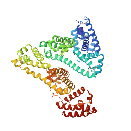
Crystal Structures of Human Serum Albumin Complexed with Monounsaturated and Polyunsaturated Fatty Acids.

Petitpas, I.Gruene, T.Bhattacharya, A.A.Curry, S.

(2001) J Mol Biol 314: 955

  • DOI: https://doi.org/10.1006/jmbi.2000.5208
  • Primary Citation of Related Structures:  
    1GNI, 1GNJ

  • PubMed Abstract: 

    The primary ligands of human serum albumin (HSA), an abundant plasma protein, are non-esterified fatty acids. In vivo, the majority of fatty acids associated with the protein are unsaturated. We present here the first high-resolution crystal structures of HSA complexed with two important unsaturated fatty acids, the monounsaturated oleic acid (C18:1) and the polyunsaturated arachidonic acid (C20:4). Both compounds are observed to occupy the seven binding sites distributed across the protein that are also bound by medium and long-chain saturated fatty acids. Although C18:1 fatty acid binds each site on HSA in a conformation almost identical with that of the corresponding saturated compound (C18:0), the presence of multiple cis double bonds in C20:4 induces distinct binding configurations at some sites. The observed restriction on binding configurations plausibly accounts for differences in the pattern of binding affinities for the primary sites between polyunsaturated fatty acids and their saturated or monounsaturated counterparts.


  • Organizational Affiliation

    Biophysics Section, Department of Biological Sciences, Blackett Laboratory, Imperial College of Science, Technology and Medicine, London SW7 2BW, UK.


Macromolecules
Find similar proteins by:  (by identity cutoff)  |  3D Structure
Entity ID: 1
MoleculeChains Sequence LengthOrganismDetailsImage
SERUM ALBUMIN585Homo sapiensMutation(s): 0 
UniProt & NIH Common Fund Data Resources
Find proteins for P02768 (Homo sapiens)
Explore P02768 
Go to UniProtKB:  P02768
PHAROS:  P02768
GTEx:  ENSG00000163631 
Entity Groups  
Sequence Clusters30% Identity50% Identity70% Identity90% Identity95% Identity100% Identity
UniProt GroupP02768
Sequence Annotations
Expand
  • Reference Sequence
Experimental Data & Validation

Experimental Data

  • Method: X-RAY DIFFRACTION
  • Resolution: 2.40 Å
  • R-Value Free: 0.242 
  • R-Value Work: 0.209 
  • R-Value Observed: 0.209 
  • Space Group: C 1 2 1
Unit Cell:
Length ( Å )Angle ( ˚ )
a = 188.386α = 90
b = 38.923β = 104.86
c = 95.477γ = 90
Software Package:
Software NamePurpose
CNSrefinement
MOSFLMdata reduction
CCP4data scaling
CNSphasing

Structure Validation

View Full Validation Report


Ligand Structure Quality Assessment 


Entry History 

Deposition Data

Revision History  (Full details and data files)

  • Version 1.0: 2002-01-01
    Type: Initial release
  • Version 1.1: 2011-05-08
    Changes: Version format compliance
  • Version 1.2: 2011-07-13
    Changes: Version format compliance
  • Version 1.3: 2019-07-24
    Changes: Data collection
  • Version 1.4: 2023-12-13
    Changes: Data collection, Database references, Other, Refinement description