1GKT

Neutron Laue diffraction structure of endothiapepsin complexed with transition state analogue inhibitor H261


Experimental Data Snapshot

  • Method: NEUTRON DIFFRACTION
  • Resolution: 2.10 Å
  • R-Value Free: 0.274 
  • R-Value Observed: 0.235 

wwPDB Validation   3D Report Full Report


This is version 2.0 of the entry. See complete history


Literature

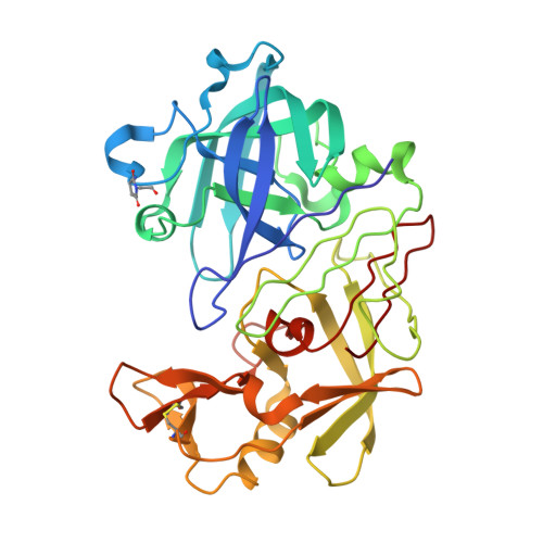
A Neutron Laue Diffraction Study of Endothiapepsin: Implications for the Aspartic Proteinase Mechanism

Coates, L.Erskine, P.T.Wood, S.P.Myles, D.A.A.Cooper, J.B.

(2001) Biochemistry 40: 13149

  • DOI: https://doi.org/10.1021/bi010626h
  • Primary Citation of Related Structures:  
    1GKT

  • PubMed Abstract: 

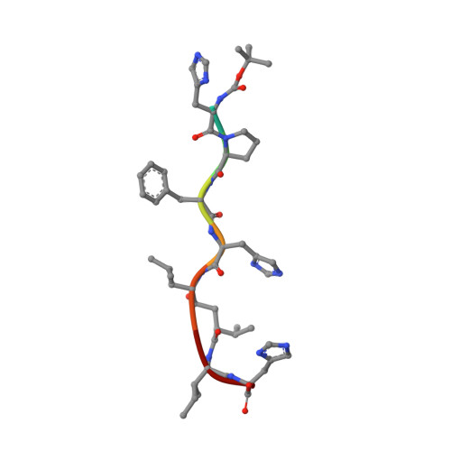
    Current proposals for the catalytic mechanism of aspartic proteinases are largely based on X-ray structures of bound oligopeptide inhibitors possessing nonhydrolyzable analogues of the scissile peptide bond. However, the positions of protons on the catalytic aspartates and the ligand in these complexes have not been determined with certainty. Thus, our objective was to locate crucial protons at the active site of an inhibitor complex since this will have major implications for a detailed understanding of the mechanism of action. We have demonstrated that high-resolution neutron diffraction data can be collected from crystals of the fungal aspartic proteinase endothiapepsin bound to a transition state analogue (H261). The neutron structure of the complex has been refined at a resolution of 2.1 A to an R-factor of 23.5% and an R(free) of 27.4%. This work represents the largest protein structure studied to date by neutron crystallography at high resolution. The neutron data demonstrate that 49% of the main chain nitrogens have exchanged their hydrogen atoms with D2O in the mother liquor. The majority of residues resisting exchange are buried within core beta-sheet regions of the molecule. The neutron maps confirm that the protein has a number of buried ionized carboxylate groups which are likely to give the molecule a net negative charge even at very low pH, thereby accounting for its low pI. The functional groups at the catalytic center have clearly undergone H-D exchange despite being buried by the inhibitor occupying the active site cleft. Most importantly, the data provide convincing evidence that Asp 215 is protonated and that Asp 32 is the negatively charged residue in the transition state complex. This has an important bearing on mechanistic proposals for this class of proteinase.


  • Organizational Affiliation

    Division of Biochemistry and Molecular Biology, School of Biological Sciences, University of Southampton, Southampton SO16 7PX, UK.


Macromolecules
Find similar proteins by:  (by identity cutoff)  |  3D Structure
Entity ID: 1
MoleculeChains Sequence LengthOrganismDetailsImage
ENDOTHIAPEPSIN329Cryphonectria parasiticaMutation(s): 0 
EC: 3.4.23.22
UniProt
Find proteins for P11838 (Cryphonectria parasitica)
Explore P11838 
Go to UniProtKB:  P11838
Entity Groups  
Sequence Clusters30% Identity50% Identity70% Identity90% Identity95% Identity100% Identity
UniProt GroupP11838
Sequence Annotations
Expand
  • Reference Sequence

Find similar proteins by:  Sequence   |   3D Structure  

Entity ID: 2
MoleculeChains Sequence LengthOrganismDetailsImage
INHIBITOR, H2618synthetic constructMutation(s): 0 
Sequence Annotations
Expand
  • Reference Sequence
Small Molecules
Modified Residues  1 Unique
IDChains TypeFormula2D DiagramParent
SUI
Query on SUI
A
L-PEPTIDE LINKINGC6 H8 N2 O4ASP, GLY
Biologically Interesting Molecules (External Reference) 1 Unique
Entity ID: 2
IDChains NameType/Class2D Diagram3D Interactions
PRD_000267
Query on PRD_000267
B
H-261 OligopeptidePeptide-like / Inhibitor
Experimental Data & Validation

Experimental Data

  • Method: NEUTRON DIFFRACTION
  • Resolution: 2.10 Å
  • R-Value Free: 0.274 
  • R-Value Observed: 0.235 
  • Space Group: P 1 21 1
Unit Cell:
Length ( Å )Angle ( ˚ )
a = 43.1α = 90
b = 75.7β = 97
c = 42.9γ = 90
Software Package:
Software NamePurpose
SHELXL-97refinement
LAUEGENdata reduction
SCALAdata scaling

Structure Validation

View Full Validation Report


Entry History 

Deposition Data

Revision History  (Full details and data files)

  • Version 1.0: 2001-11-20
    Type: Initial release
  • Version 1.1: 2011-07-13
    Changes: Atomic model, Database references, Derived calculations, Non-polymer description, Structure summary, Version format compliance
  • Version 1.2: 2012-11-30
    Changes: Other
  • Version 1.3: 2013-01-16
    Changes: Atomic model, Other
  • Version 1.4: 2013-03-06
    Changes: Other
  • Version 1.5: 2017-03-22
    Changes: Atomic model, Data collection
  • Version 1.6: 2018-02-14
    Changes: Data collection
  • Version 2.0: 2023-11-15
    Changes: Atomic model, Data collection, Database references, Derived calculations, Other