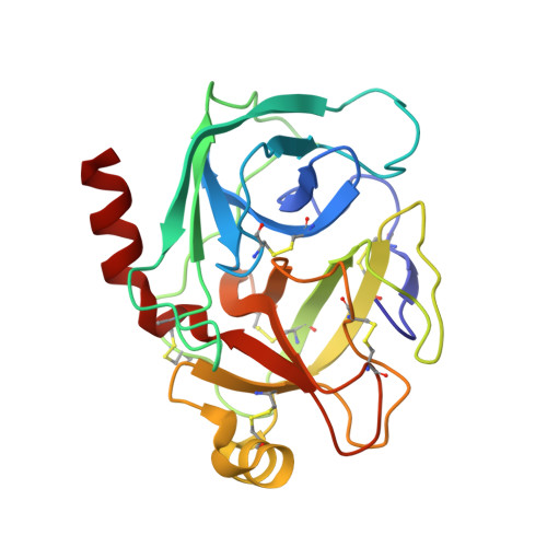
Structure of an acyl-enzyme intermediate during catalysis: (guanidinobenzoyl)trypsin.
Mangel, W.F., Singer, P.T., Cyr, D.M., Umland, T.C., Toledo, D.L., Stroud, R.M., Pflugrath, J.W., Sweet, R.M.(1990) Biochemistry 29: 8351-8357
- PubMed: 2252895
- DOI: https://doi.org/10.1021/bi00488a022
- Primary Citation of Related Structures:
1GBT - PubMed Abstract:
The crystal and molecular structure of trypsin at a transiently stable intermediate step during catalysis has been determined by X-ray diffraction methods. Bovine trypsin cleaved the substrate p-nitrophenyl p-guanidinobenzoate during crystallization under conditions in which the acyl-enzyme intermediate, (guanidinobenzoyl)trypsin, was stable. Orthorhombic crystals formed in space group P2(1)2(1)2(1), with a = 63.74, b = 63.54, and c = 68.93 A. This is a crystal form of bovine trypsin for which a molecular structure has not been reported. Diffraction data were measured with a FAST (Enraf Nonius) diffractometer. The structure was refined to a crystallographic residual of R = 0.16 for data in the resolution range 7.0-2.0 A. The refined model of (guanidinobenzoyl)trypsin provides insight into the structural basis for its slow rate of deacylation, which in solution at 25 degrees C and pH 7.4 exhibits a t1/2 of 12 h. In addition to the rotation of the Ser-195 hydroxyl away from His-157, C beta of Ser-195 moves 0.7 A toward Asp-189 at the bottom of the active site, with respect to the native structure. This allows formation of energetically favorable H bonds and an ion pair between the carboxylate of Asp-189 and the guanidino group of the substrate. This movement is dictated by the rigidity of the aromatic ring in guanidinobenzoate--model-building indicates that this should not occur when arginine, with its more flexible aliphatic backbone, forms the ester bond with Ser-195. As a consequence, highly ordered water molecules in the active site are no longer close enough to the scissile ester bond to serve as potential nucleophiles for hydrolysis.(ABSTRACT TRUNCATED AT 250 WORDS)
Organizational Affiliation:
Biology Department, Brookhaven National Laboratory, Upton, New York 11973.