1G2N

CRYSTAL STRUCTURE OF THE LIGAND BINDING DOMAIN OF THE ULTRASPIRACLE PROTEIN USP, THE ORTHOLOG OF RXRS IN INSECTS


Experimental Data Snapshot

  • Method: X-RAY DIFFRACTION
  • Resolution: 1.65 Å
  • R-Value Free: 0.244 
  • R-Value Work: 0.210 
  • R-Value Observed: 0.210 

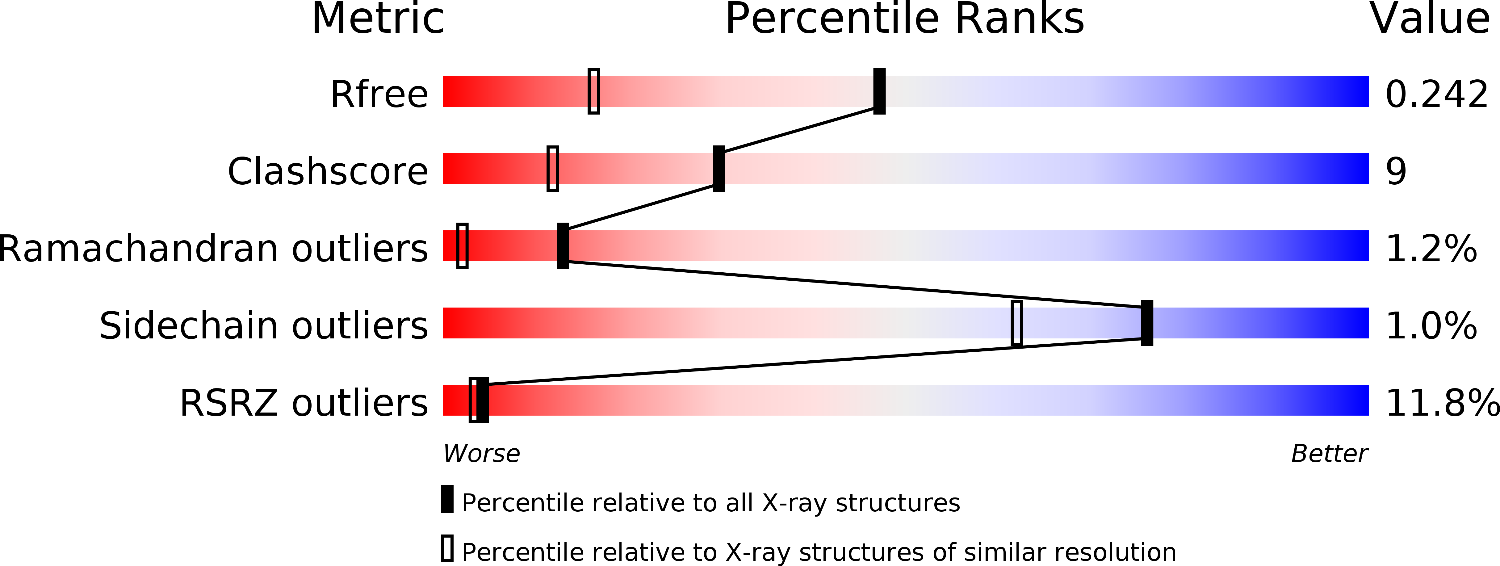
wwPDB Validation   3D Report Full Report


Ligand Structure Quality Assessment 


This is version 1.3 of the entry. See complete history


Literature

Crystal structure of the ligand-binding domain of the ultraspiracle protein USP, the ortholog of retinoid X receptors in insects.

Billas, I.M.Moulinier, L.Rochel, N.Moras, D.

(2001) J Biol Chem 276: 7465-7474

  • DOI: https://doi.org/10.1074/jbc.M008926200
  • Primary Citation of Related Structures:  
    1G2N

  • PubMed Abstract: 

    The major postembryonic developmental events happening in insect life, including molting and metamorphosis, are regulated and coordinated temporally by pulses of ecdysone. The biological activity of this steroid hormone is mediated by two nuclear receptors: the ecdysone receptor (EcR) and the Ultraspiracle protein (USP). The crystal structure of the ligand-binding domain from the lepidopteran Heliothis virescens USP reported here shows that the loop connecting helices H1 and H3 precludes the canonical agonist conformation. The key residues that stabilize this unique loop conformation are strictly conserved within the lepidopteran USP family. The presence of an unexpected bound ligand that drives an unusual antagonist conformation confirms the induced-fit mechanism accompanying the ligand binding. The ligand-binding pocket exhibits a retinoid X receptor-like anchoring part near a conserved arginine, which could interact with a USP ligand functional group. The structure of this receptor provides the template for designing inhibitors, which could be utilized as a novel type of environmentally safe insecticides.


  • Organizational Affiliation

    Genomics and Structural Biology Laboratory, UPR 9004, Institut de Génétique et de Biologie Moléculaire et Cellulaire, CNRS/INSERM/Université Louis Pasteur, 1 rue Laurent Fries, 67404 Illkirch Cedex, Cité Universitaire de Strasbourg, France.


Macromolecules
Find similar proteins by:  (by identity cutoff)  |  3D Structure
Entity ID: 1
MoleculeChains Sequence LengthOrganismDetailsImage
ULTRASPIRACLE PROTEIN264Heliothis virescensMutation(s): 0 
Gene Names: USP LOCUS
UniProt
Find proteins for Q7SIF6 (Heliothis virescens)
Explore Q7SIF6 
Go to UniProtKB:  Q7SIF6
Entity Groups  
Sequence Clusters30% Identity50% Identity70% Identity90% Identity95% Identity100% Identity
UniProt GroupQ7SIF6
Sequence Annotations
Expand
  • Reference Sequence
Small Molecules
Ligands 1 Unique
IDChains Name / Formula / InChI Key2D Diagram3D Interactions
EPH
Query on EPH

Download Ideal Coordinates CCD File 
B [auth A]L-ALPHA-PHOSPHATIDYL-BETA-OLEOYL-GAMMA-PALMITOYL-PHOSPHATIDYLETHANOLAMINE
C39 H68 N O8 P
MABRTXOVHMDVAT-AAEGOEIASA-N
Experimental Data & Validation

Experimental Data

  • Method: X-RAY DIFFRACTION
  • Resolution: 1.65 Å
  • R-Value Free: 0.244 
  • R-Value Work: 0.210 
  • R-Value Observed: 0.210 
  • Space Group: P 43 2 2
Unit Cell:
Length ( Å )Angle ( ˚ )
a = 58.211α = 90
b = 58.211β = 90
c = 144.687γ = 90
Software Package:
Software NamePurpose
CNSrefinement
DENZOdata reduction
SCALEPACKdata scaling
CNSphasing

Structure Validation

View Full Validation Report


Ligand Structure Quality Assessment 


Entry History 

Deposition Data

Revision History  (Full details and data files)

  • Version 1.0: 2001-04-21
    Type: Initial release
  • Version 1.1: 2008-04-27
    Changes: Version format compliance
  • Version 1.2: 2011-07-13
    Changes: Version format compliance
  • Version 1.3: 2024-02-07
    Changes: Data collection, Database references, Derived calculations