1FYF

CRYSTAL STRUCTURE OF A TRUNCATED FORM OF THREONYL-TRNA SYNTHETASE COMPLEXED WITH A SERYL ADENYLATE ANALOG


Experimental Data Snapshot

  • Method: X-RAY DIFFRACTION
  • Resolution: 1.65 Å
  • R-Value Free: 0.237 
  • R-Value Work: 0.218 
  • R-Value Observed: 0.218 

wwPDB Validation   3D Report Full Report


Ligand Structure Quality Assessment 


This is version 1.3 of the entry. See complete history


Literature

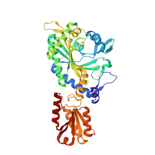
Transfer RNA-mediated editing in threonyl-tRNA synthetase. The class II solution to the double discrimination problem.

Dock-Bregeon, A.Sankaranarayanan, R.Romby, P.Caillet, J.Springer, M.Rees, B.Francklyn, C.S.Ehresmann, C.Moras, D.

(2000) Cell 103: 877-884

  • DOI: https://doi.org/10.1016/s0092-8674(00)00191-4
  • Primary Citation of Related Structures:  
    1FYF

  • PubMed Abstract: 

    Threonyl-tRNA synthetase, a class II synthetase, uses a unique zinc ion to discriminate against the isosteric valine at the activation step. The crystal structure of the enzyme with an analog of seryl adenylate shows that the noncognate serine cannot be fully discriminated at that step. We show that hydrolysis of the incorrectly formed ser-tRNA(Thr) is performed at a specific site in the N-terminal domain of the enzyme. The present study suggests that both classes of synthetases use effectively the ability of the CCA end of tRNA to switch between a hairpin and a helical conformation for aminoacylation and editing. As a consequence, the editing mechanism of both classes of synthetases can be described as mirror images, as already seen for tRNA binding and amino acid activation.


  • Organizational Affiliation

    UPR 9004 Biologie Structurale IGBMC, CNRS/INSERM/ULP BP163 67404 Cedex, Illkirch, France.


Macromolecules
Find similar proteins by:  (by identity cutoff)  |  3D Structure
Entity ID: 1
MoleculeChains Sequence LengthOrganismDetailsImage
THREONYL-TRNA SYNTHETASE
A, B
401Escherichia coliMutation(s): 0 
EC: 6.1.1.3
UniProt
Find proteins for P0A8M3 (Escherichia coli (strain K12))
Explore P0A8M3 
Go to UniProtKB:  P0A8M3
Entity Groups  
Sequence Clusters30% Identity50% Identity70% Identity90% Identity95% Identity100% Identity
UniProt GroupP0A8M3
Sequence Annotations
Expand
  • Reference Sequence
Experimental Data & Validation

Experimental Data

  • Method: X-RAY DIFFRACTION
  • Resolution: 1.65 Å
  • R-Value Free: 0.237 
  • R-Value Work: 0.218 
  • R-Value Observed: 0.218 
  • Space Group: P 21 21 21
Unit Cell:
Length ( Å )Angle ( ˚ )
a = 86.95α = 90
b = 109.52β = 90
c = 115.37γ = 90
Software Package:
Software NamePurpose
AMoREphasing
CNSrefinement
DENZOdata reduction
SCALEPACKdata scaling

Structure Validation

View Full Validation Report


Ligand Structure Quality Assessment 


Entry History 

Deposition Data

Revision History  (Full details and data files)

  • Version 1.0: 2000-12-27
    Type: Initial release
  • Version 1.1: 2008-04-27
    Changes: Version format compliance
  • Version 1.2: 2011-07-13
    Changes: Version format compliance
  • Version 1.3: 2024-02-07
    Changes: Data collection, Database references, Derived calculations