1FUI

L-FUCOSE ISOMERASE FROM ESCHERICHIA COLI


Experimental Data Snapshot

  • Method: X-RAY DIFFRACTION
  • Resolution: 2.50 Å
  • R-Value Free: 0.209 
  • R-Value Work: 0.162 
  • R-Value Observed: 0.162 

wwPDB Validation   3D Report Full Report


Ligand Structure Quality Assessment 


This is version 1.3 of the entry. See complete history


Literature

Structure and mechanism of L-fucose isomerase from Escherichia coli.

Seemann, J.E.Schulz, G.E.

(1997) J Mol Biol 273: 256-268

  • DOI: https://doi.org/10.1006/jmbi.1997.1280
  • Primary Citation of Related Structures:  
    1FUI

  • PubMed Abstract: 

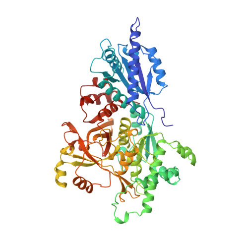
    The three-dimensional structure of L-fucose isomerase from Escherichia coli has been determined by X-ray crystallography at 2.5 A resolution. This ketol isomerase converts the aldose L-fucose into the corresponding ketose L-fuculose using Mn2+ as a cofactor. Being a hexamer with 64,976 Da per subunit, L-fucose isomerase is the largest structurally known ketol isomerase. The enzyme shows neither sequence nor structural similarity with other ketol isomerases. The hexamer obeys D3 symmetry and forms the crystallographic asymmetric unit. The strict and favorably oriented local symmetry allowed for a computational phase extension from 7.3 A to 2.5 A resolution. The structure was solved with an L-fucitol molecule bound to the catalytic center such that the hydroxyl groups at positions 1 and 2 are ligands of the manganese ion. Most likely, L-fucitol mimics a bound L-fucose molecule in its open chain form. The protein environment suggests strongly that the reaction belongs to the ene-diol type.


  • Organizational Affiliation

    Institut für Organische Chemie und Biochemie, Albert-Ludwigs-Universität, Freiburg im Breisgau, Germany.


Macromolecules
Find similar proteins by:  (by identity cutoff)  |  3D Structure
Entity ID: 1
MoleculeChains Sequence LengthOrganismDetailsImage
L-FUCOSE ISOMERASE
A, B, C, D, E
A, B, C, D, E, F
591Escherichia coli K-12Mutation(s): 0 
Gene Names: FUCI
EC: 5.3.1.3
UniProt
Find proteins for P69922 (Escherichia coli (strain K12))
Explore P69922 
Go to UniProtKB:  P69922
Entity Groups  
Sequence Clusters30% Identity50% Identity70% Identity90% Identity95% Identity100% Identity
UniProt GroupP69922
Sequence Annotations
Expand
  • Reference Sequence
Small Molecules
Ligands 3 Unique
IDChains Name / Formula / InChI Key2D Diagram3D Interactions
FOC
Query on FOC

Download Ideal Coordinates CCD File 
I [auth A]
K [auth B]
M [auth C]
P [auth D]
R [auth E]
I [auth A],
K [auth B],
M [auth C],
P [auth D],
R [auth E],
S [auth F]
FUCITOL
C6 H14 O5
SKCKOFZKJLZSFA-KCDKBNATSA-N
SO4
Query on SO4

Download Ideal Coordinates CCD File 
H [auth A],
O [auth D]
SULFATE ION
O4 S
QAOWNCQODCNURD-UHFFFAOYSA-L
MN
Query on MN

Download Ideal Coordinates CCD File 
G [auth A],
J [auth B],
L [auth C],
N [auth D],
Q [auth E]
MANGANESE (II) ION
Mn
WAEMQWOKJMHJLA-UHFFFAOYSA-N
Experimental Data & Validation

Experimental Data

  • Method: X-RAY DIFFRACTION
  • Resolution: 2.50 Å
  • R-Value Free: 0.209 
  • R-Value Work: 0.162 
  • R-Value Observed: 0.162 
  • Space Group: P 21 21 21
Unit Cell:
Length ( Å )Angle ( ˚ )
a = 127.3α = 90
b = 128.3β = 90
c = 239.4γ = 90
Software Package:
Software NamePurpose
X-PLORmodel building
X-PLORrefinement
DENZOdata reduction
CCP4data scaling
X-PLORphasing

Structure Validation

View Full Validation Report


Ligand Structure Quality Assessment 


Entry History 

Deposition Data

Revision History  (Full details and data files)

  • Version 1.0: 1997-10-15
    Type: Initial release
  • Version 1.1: 2008-03-03
    Changes: Version format compliance
  • Version 1.2: 2011-07-13
    Changes: Source and taxonomy, Version format compliance
  • Version 1.3: 2024-02-07
    Changes: Data collection, Database references, Derived calculations, Other