1FS0

COMPLEX OF GAMMA/EPSILON ATP SYNTHASE FROM E.COLI


Experimental Data Snapshot

  • Method: X-RAY DIFFRACTION
  • Resolution: 2.10 Å
  • R-Value Free: 0.269 
  • R-Value Work: 0.216 
  • R-Value Observed: 0.227 

wwPDB Validation   3D Report Full Report


This is version 1.3 of the entry. See complete history


Literature

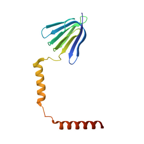
Structure of the gamma-epsilon complex of ATP synthase.

Rodgers, A.J.Wilce, M.C.

(2000) Nat Struct Biol 7: 1051-1054

  • DOI: https://doi.org/10.1038/80975
  • Primary Citation of Related Structures:  
    1FS0

  • PubMed Abstract: 

    ATP synthases (F(1)F(o)-ATPases) use energy released by the movement of protons down a transmembrane electrochemical gradient to drive the synthesis of ATP, the universal biological energy currency. Proton flow through F(o) drives rotation of a ring of c-subunits and a complex of the gamma and epsilon-subunits, causing cyclical conformational changes in F(1) that are required for catalysis. The crystal structure of a large portion of F(1) has been resolved. However, the structure of the central portion of the enzyme, through which conformational changes in F(o) are communicated to F(1), has until now remained elusive. Here we report the crystal structure of a complex of the epsilon-subunit and the central domain of the gamma-subunit refined at 2.1 A resolution. The structure reveals how rotation of these subunits causes large conformational changes in F(1), and thereby provides new insights into energy coupling between F(o) and F(1).


  • Organizational Affiliation

    Crystallography Centre Department of Pharmacology, University of Western Australia and Western Australian Institute for Medical Research, Nedlands Western Australia 6907, Australia.


Macromolecules
Find similar proteins by:  (by identity cutoff)  |  3D Structure
Entity ID: 1
MoleculeChains Sequence LengthOrganismDetailsImage
ATP SYNTHASE EPSILON SUBUNITA [auth E]138Escherichia coliMutation(s): 0 
UniProt
Find proteins for P0A6E6 (Escherichia coli (strain K12))
Explore P0A6E6 
Go to UniProtKB:  P0A6E6
Entity Groups  
Sequence Clusters30% Identity50% Identity70% Identity90% Identity95% Identity100% Identity
UniProt GroupP0A6E6
Sequence Annotations
Expand
  • Reference Sequence
Find similar proteins by:  (by identity cutoff)  |  3D Structure
Entity ID: 2
MoleculeChains Sequence LengthOrganismDetailsImage
ATP SYNTHASE GAMMA SUBUNITB [auth G]230Escherichia coliMutation(s): 0 
UniProt
Find proteins for P0ABA6 (Escherichia coli (strain K12))
Explore P0ABA6 
Go to UniProtKB:  P0ABA6
Entity Groups  
Sequence Clusters30% Identity50% Identity70% Identity90% Identity95% Identity100% Identity
UniProt GroupP0ABA6
Sequence Annotations
Expand
  • Reference Sequence
Experimental Data & Validation

Experimental Data

  • Method: X-RAY DIFFRACTION
  • Resolution: 2.10 Å
  • R-Value Free: 0.269 
  • R-Value Work: 0.216 
  • R-Value Observed: 0.227 
  • Space Group: C 2 2 21
Unit Cell:
Length ( Å )Angle ( ˚ )
a = 76.72α = 90
b = 176.09β = 90
c = 67.07γ = 90
Software Package:
Software NamePurpose
CNSrefinement
DENZOdata reduction
SCALEPACKdata scaling
CNSphasing

Structure Validation

View Full Validation Report


Entry History 

Deposition Data

Revision History  (Full details and data files)

  • Version 1.0: 2001-05-01
    Type: Initial release
  • Version 1.1: 2008-04-27
    Changes: Version format compliance
  • Version 1.2: 2011-07-13
    Changes: Version format compliance
  • Version 1.3: 2024-02-07
    Changes: Data collection, Database references